A postgraduate student once came to us with a straightforward question: which microscope should her department buy so students could see the coronavirus?
The honest answer was none of them.
Not because the budget was small, but because no optical microscope on earth can show a virus. Light itself is too big. And that surprises almost everyone the first time they hear it.
That single limitation is what created the electron microscope, and it is the real difference between the two families of instruments. Here is the full comparison, in plain terms.
What is the difference between an optical microscope and an electron microscope?
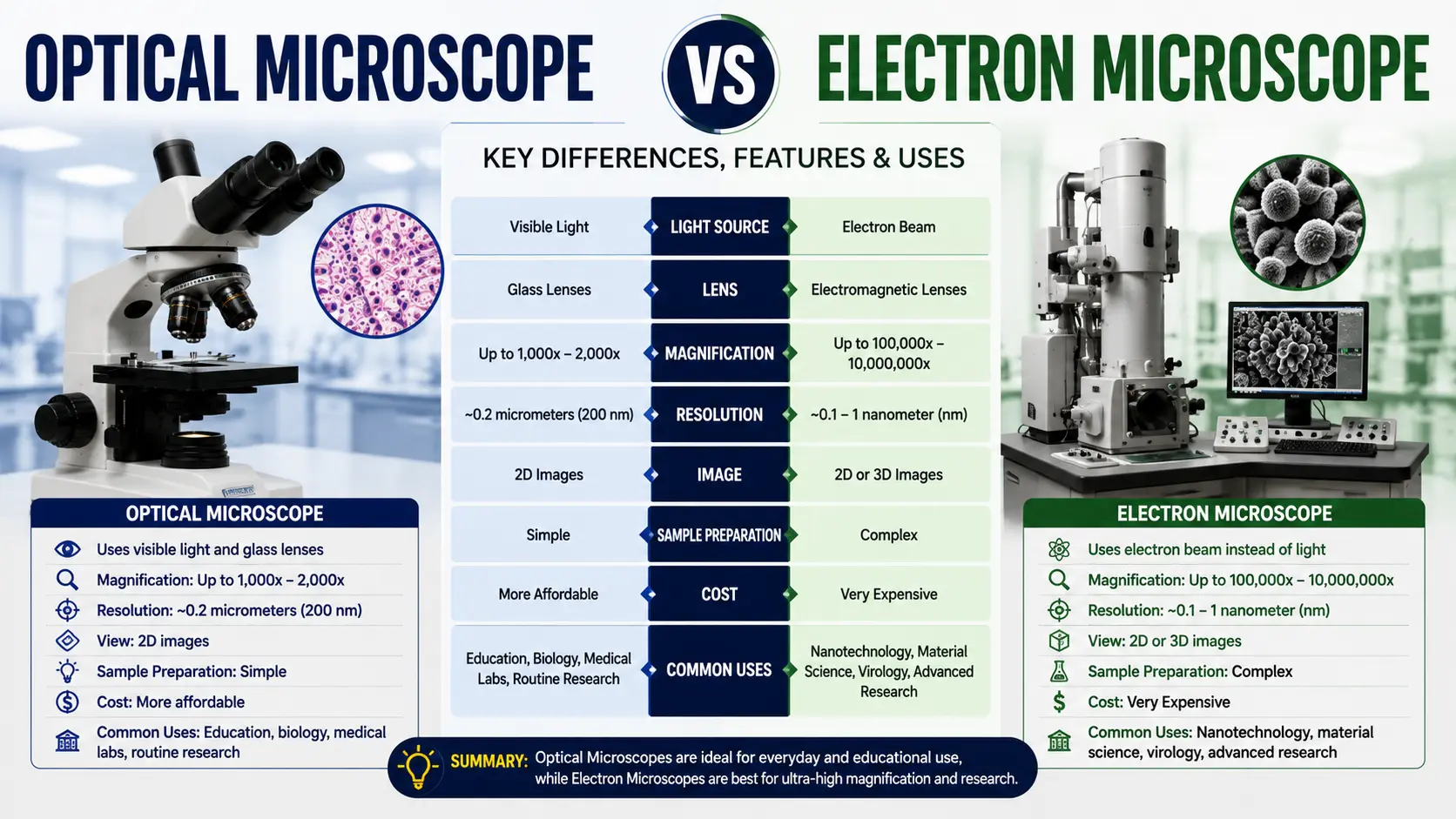
An optical microscope uses visible light and glass lenses to magnify samples up to about 1000x, showing living specimens in natural colour. An electron microscope uses a beam of electrons and electromagnetic lenses to reach over 1,00,000x with far higher resolution, but samples must be dried, coated and placed inside a vacuum.
One uses light. The other uses electrons. Everything else follows from that.
Quick Comparison Table
| Point of Difference | Optical Microscope | Electron Microscope |
| Source | Visible light (LED, halogen, mirror) | Beam of electrons |
| Lenses | Glass lenses | Electromagnetic coils |
| Maximum useful magnification | 1000x – 1500x | 1,00,000x – 10,00,000x |
| Resolution limit | About 200 nm | About 0.1 – 0.2 nm |
| Living samples | Yes | No |
| Colour | Natural colour | Greyscale only |
| Environment needed | Normal room, normal air | High vacuum chamber |
| Sample preparation | Slide and cover slip, sometimes staining | Drying, sectioning, metal coating |
| Operator training | Minimal | Specialised |
| Space required | A lab bench | A dedicated, vibration-free room |
| Price in India | ₹8,000 – ₹12 lakh | ₹40 lakh – several crore |
| Used for | Cells, bacteria, tissue, routine diagnostics | Viruses, nanoparticles, crystal structure |
What is an Optical Microscope?
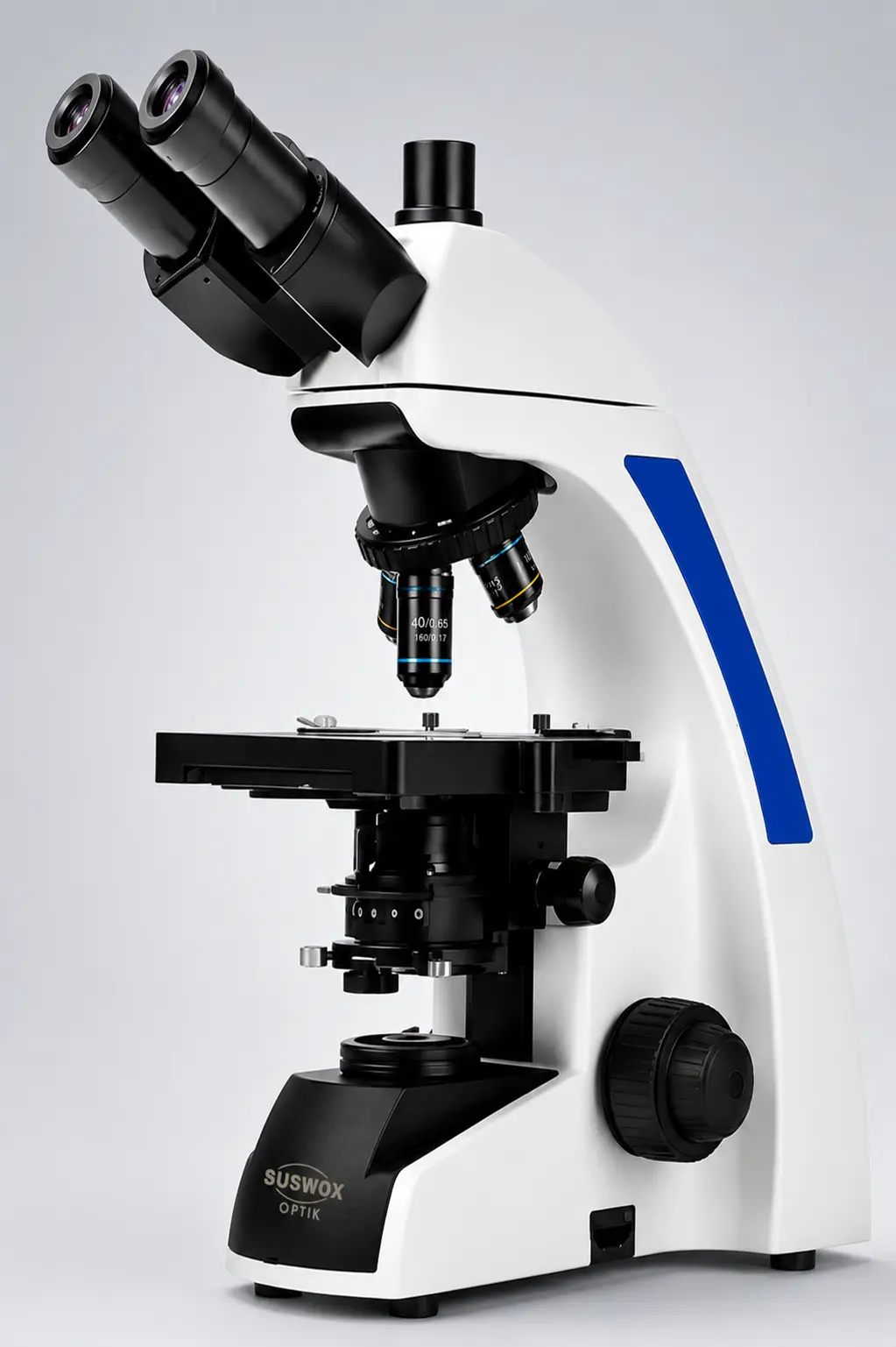
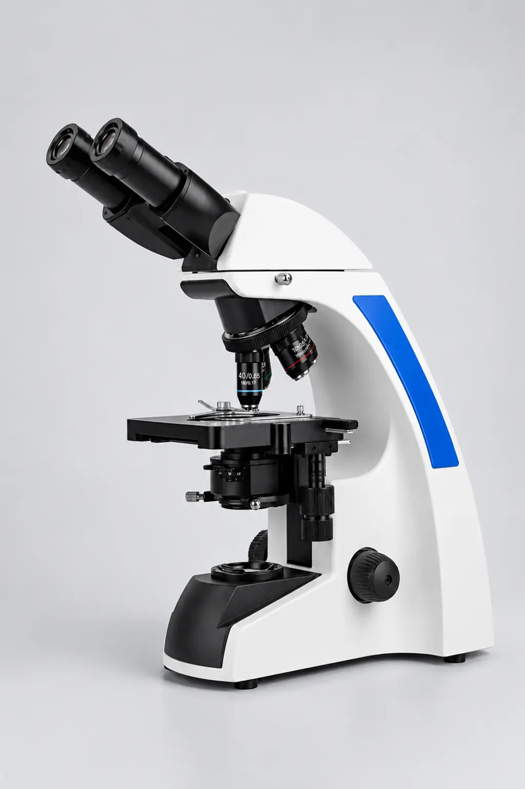
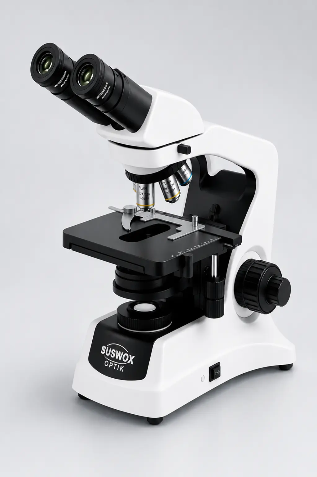
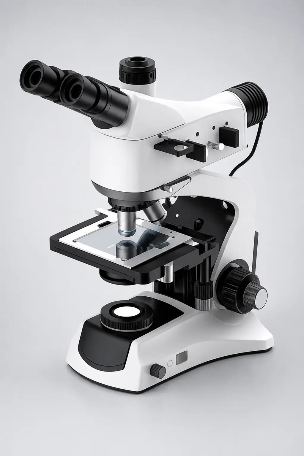
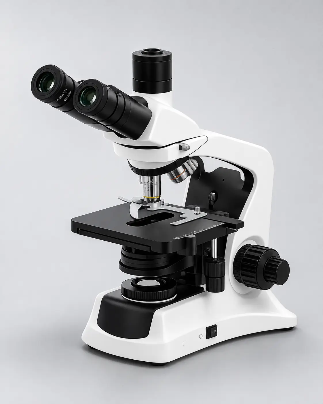
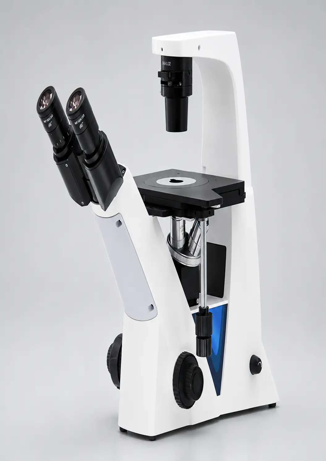
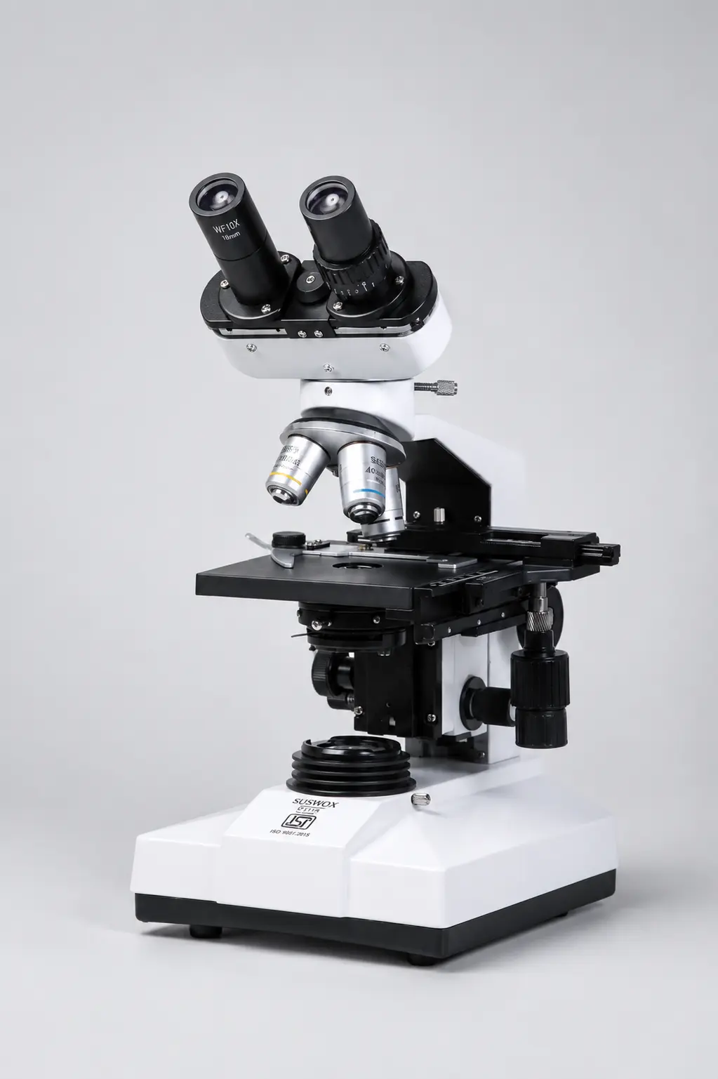
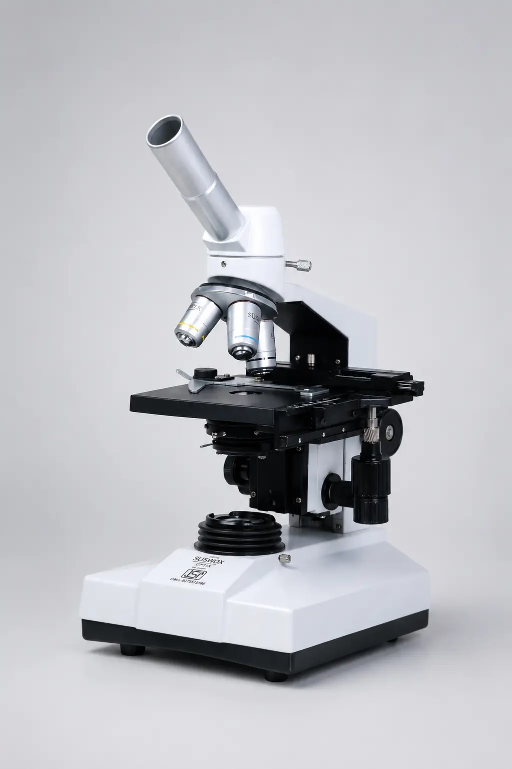
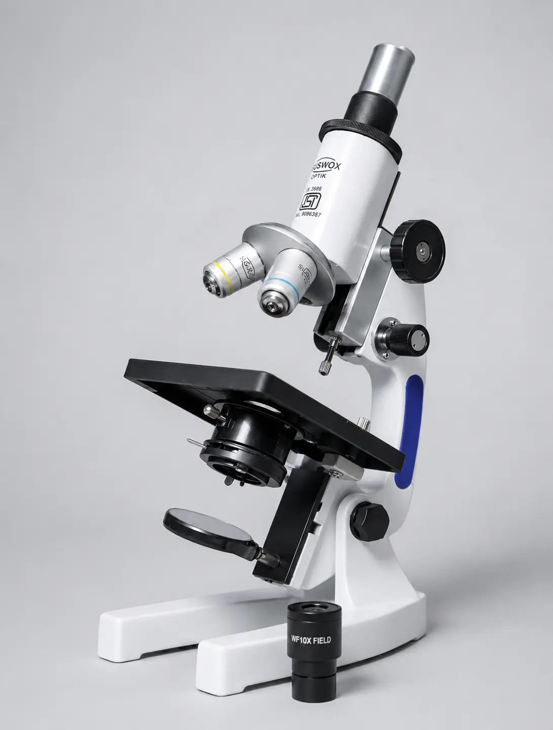
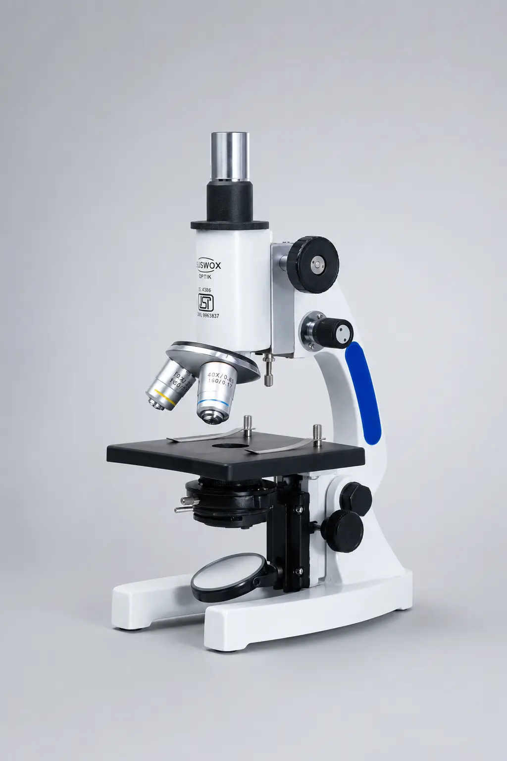
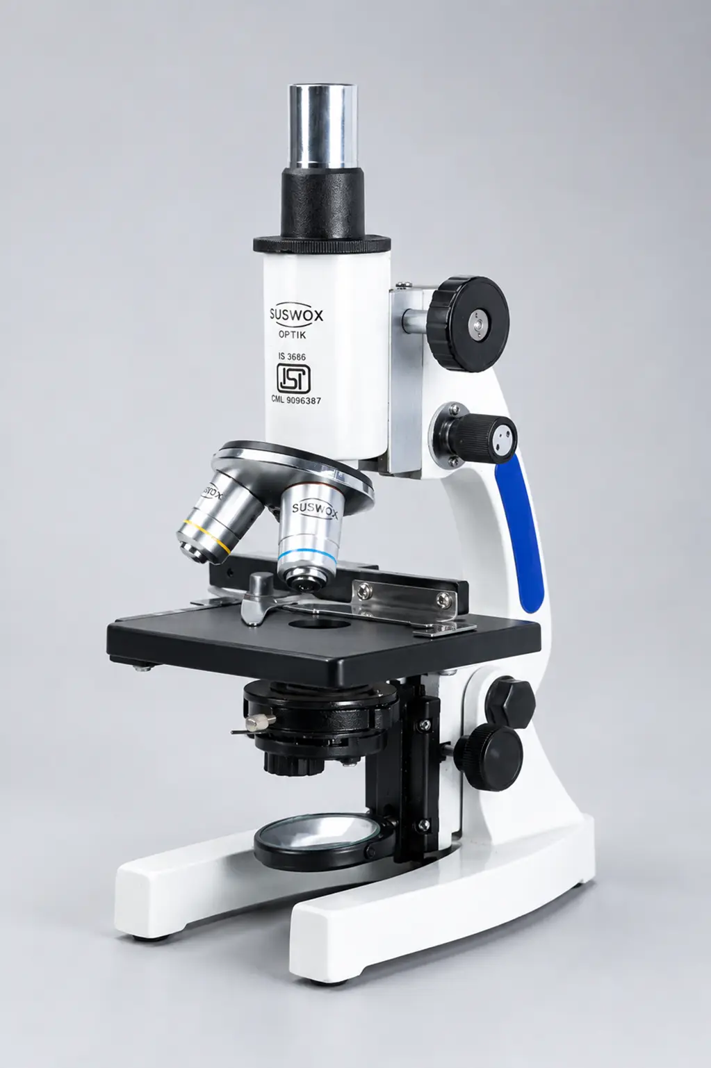
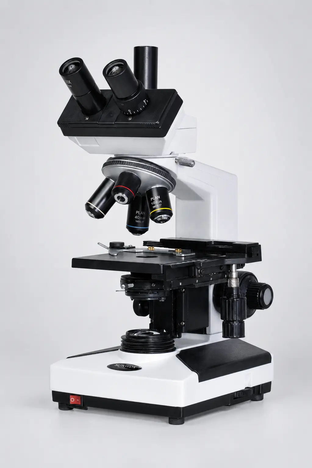
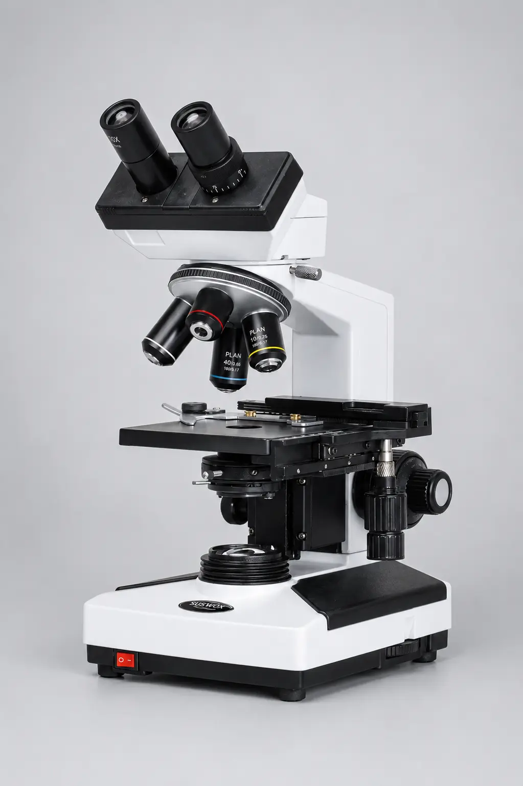
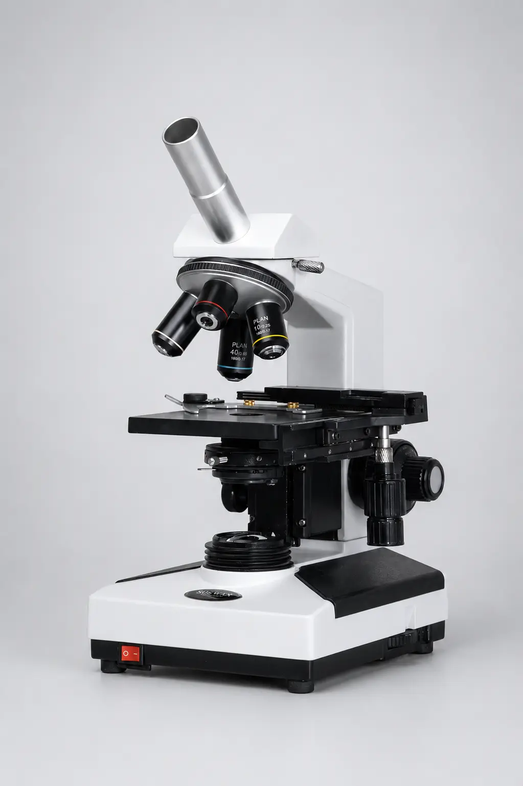
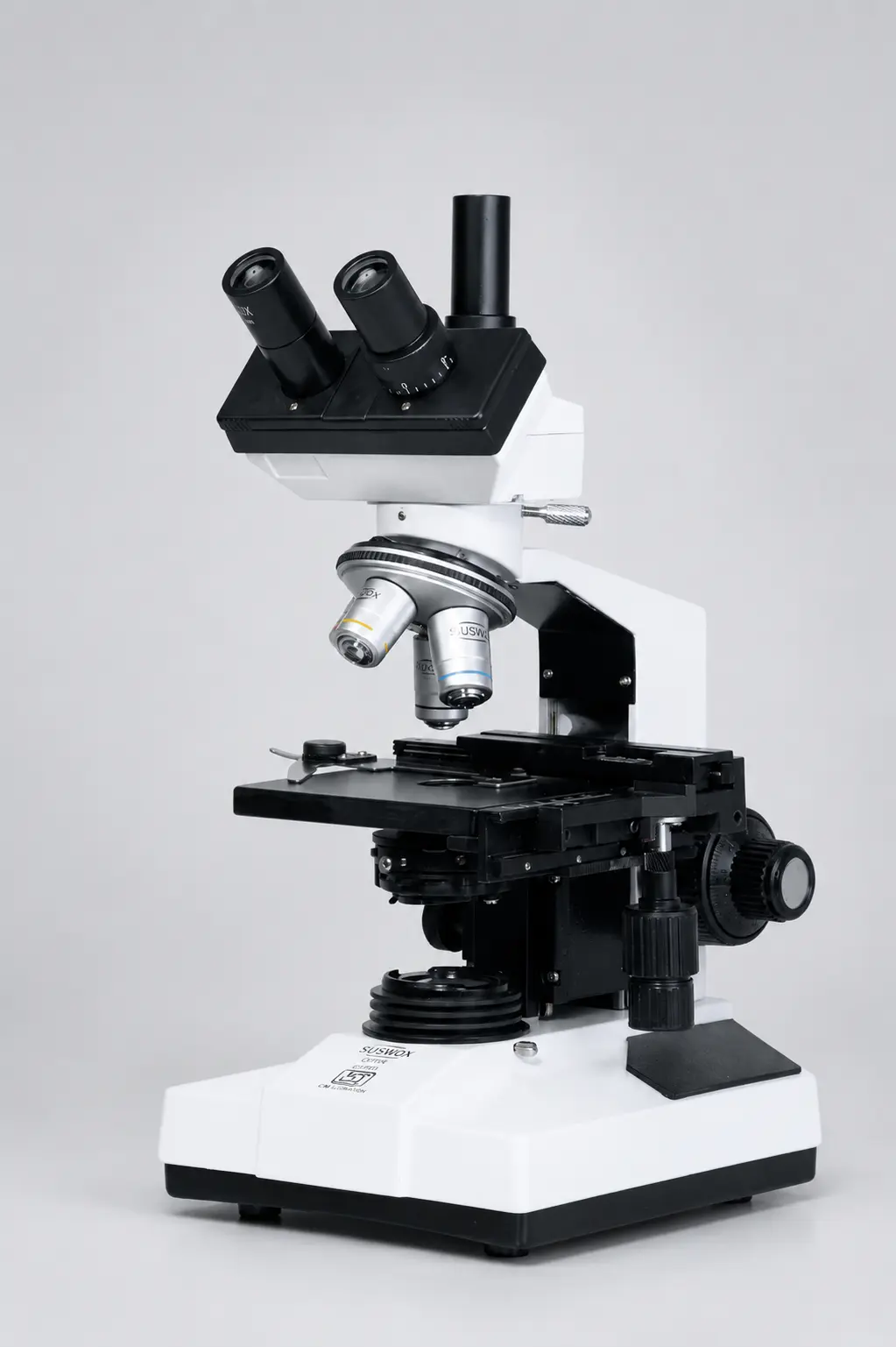
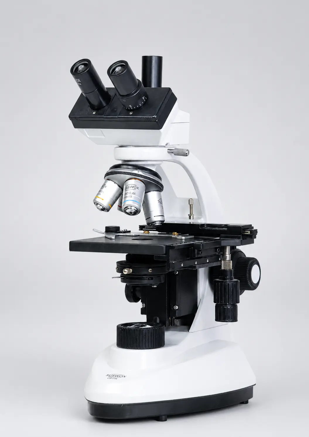
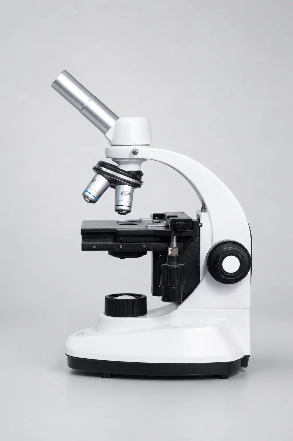
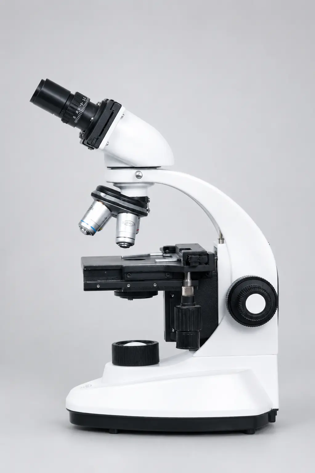
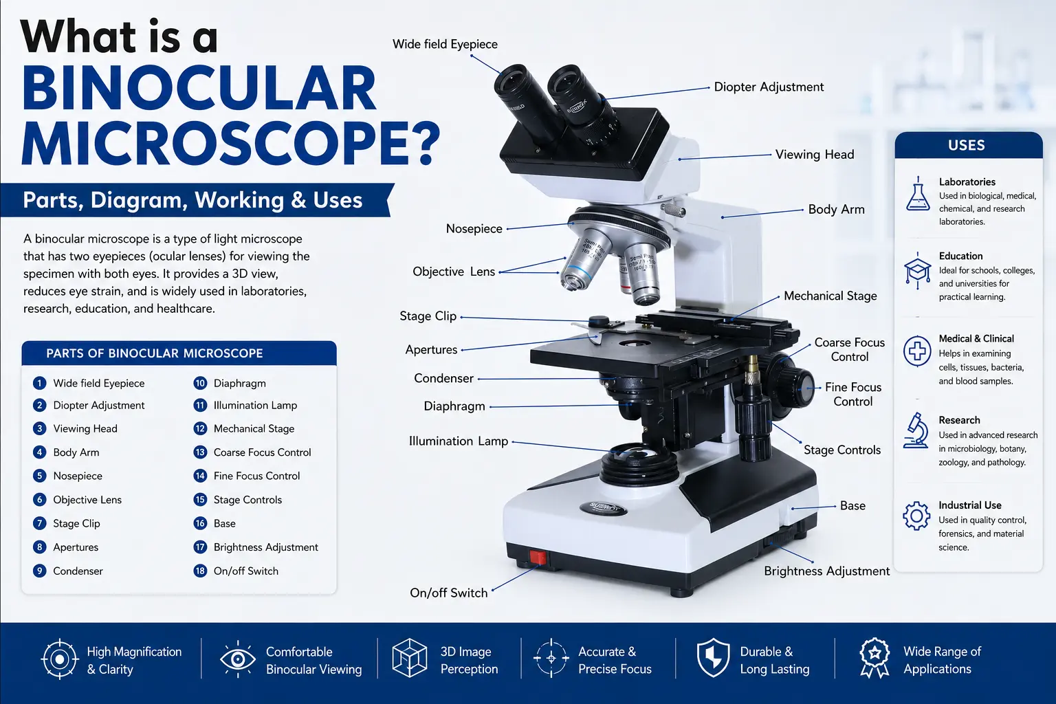
An optical microscope — also called a light microscope — magnifies objects using visible light and a system of glass lenses. This is the family that covers almost every microscope you have ever used.
It includes:
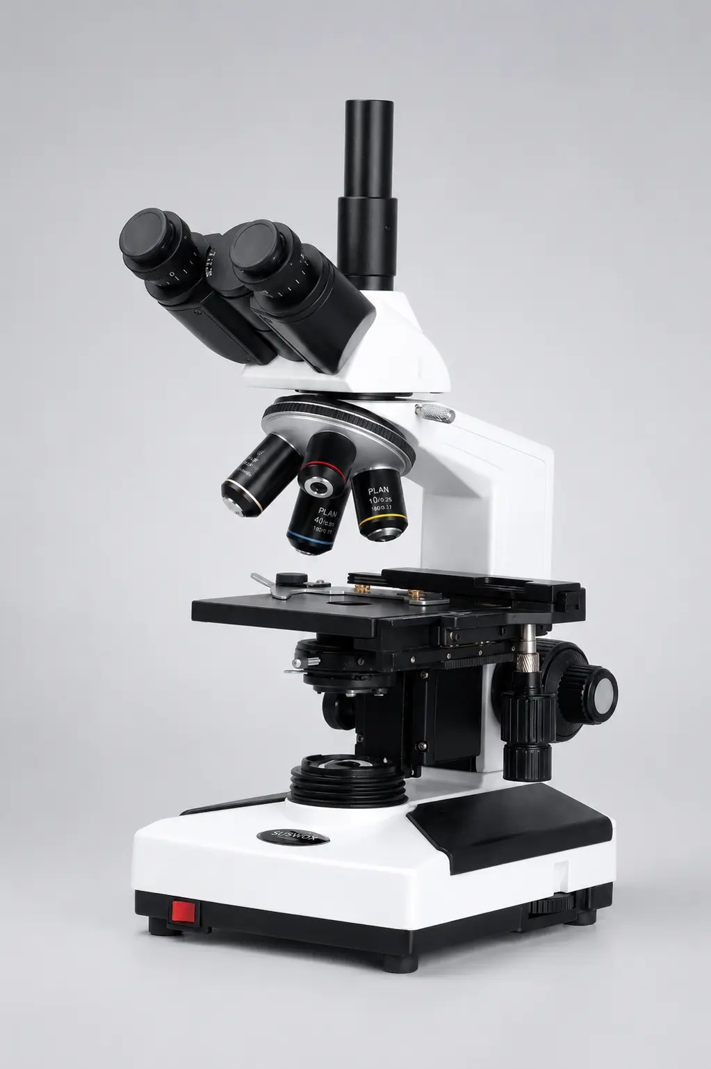
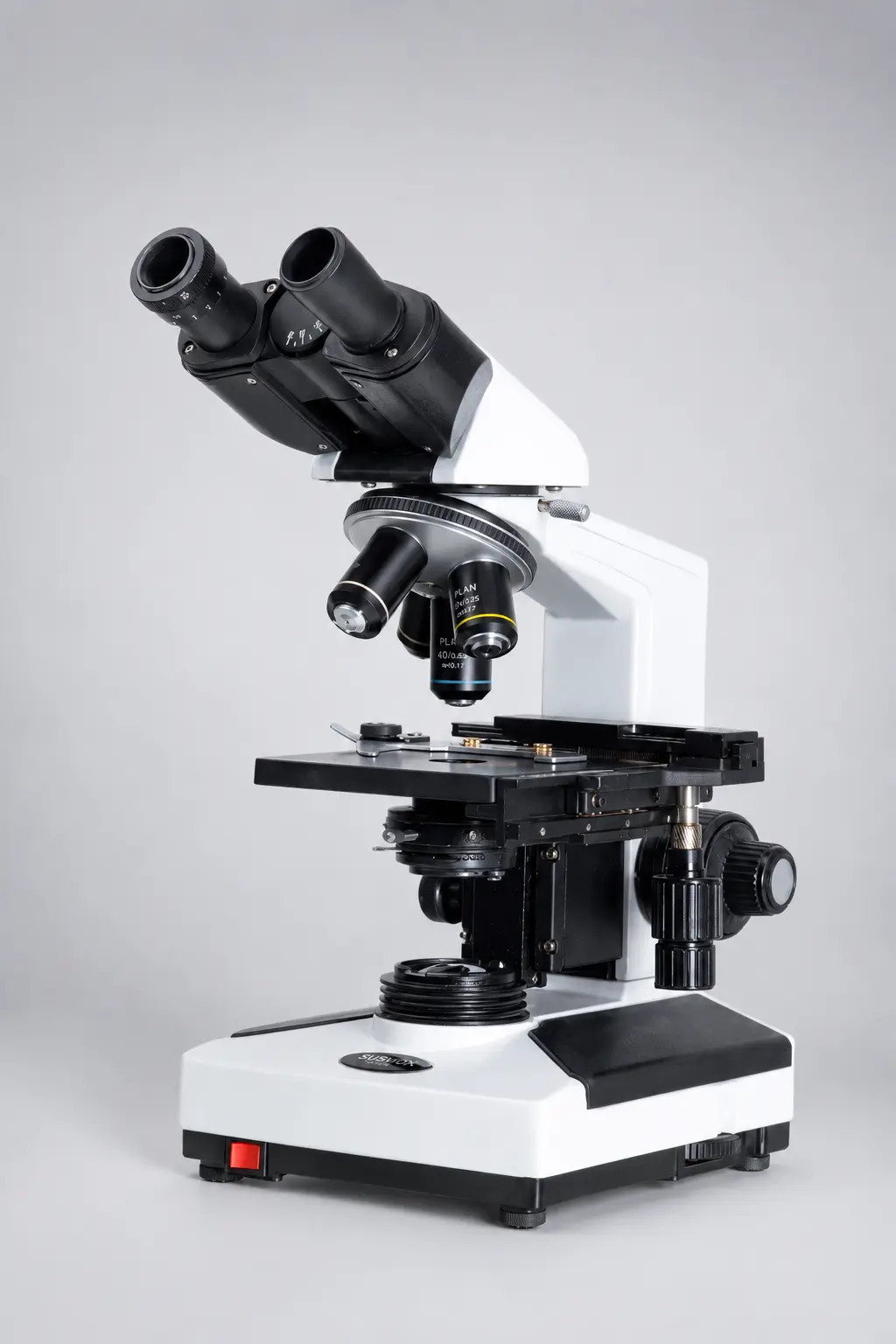
- Compound microscopes (pathology, biology, teaching)
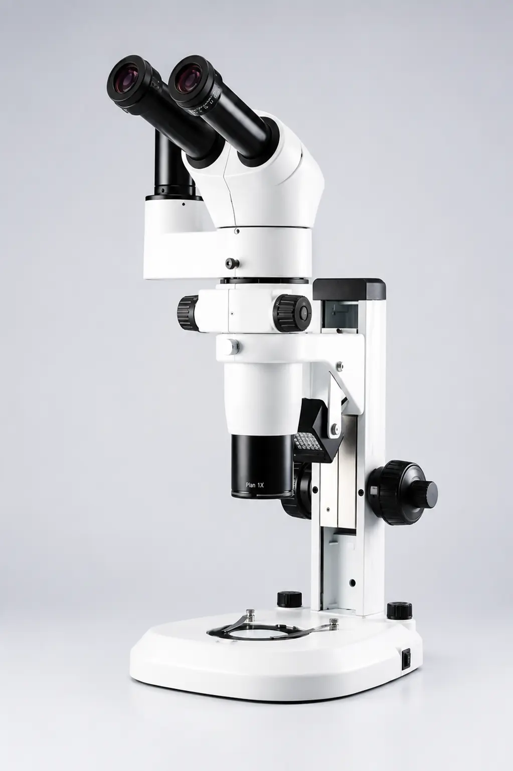
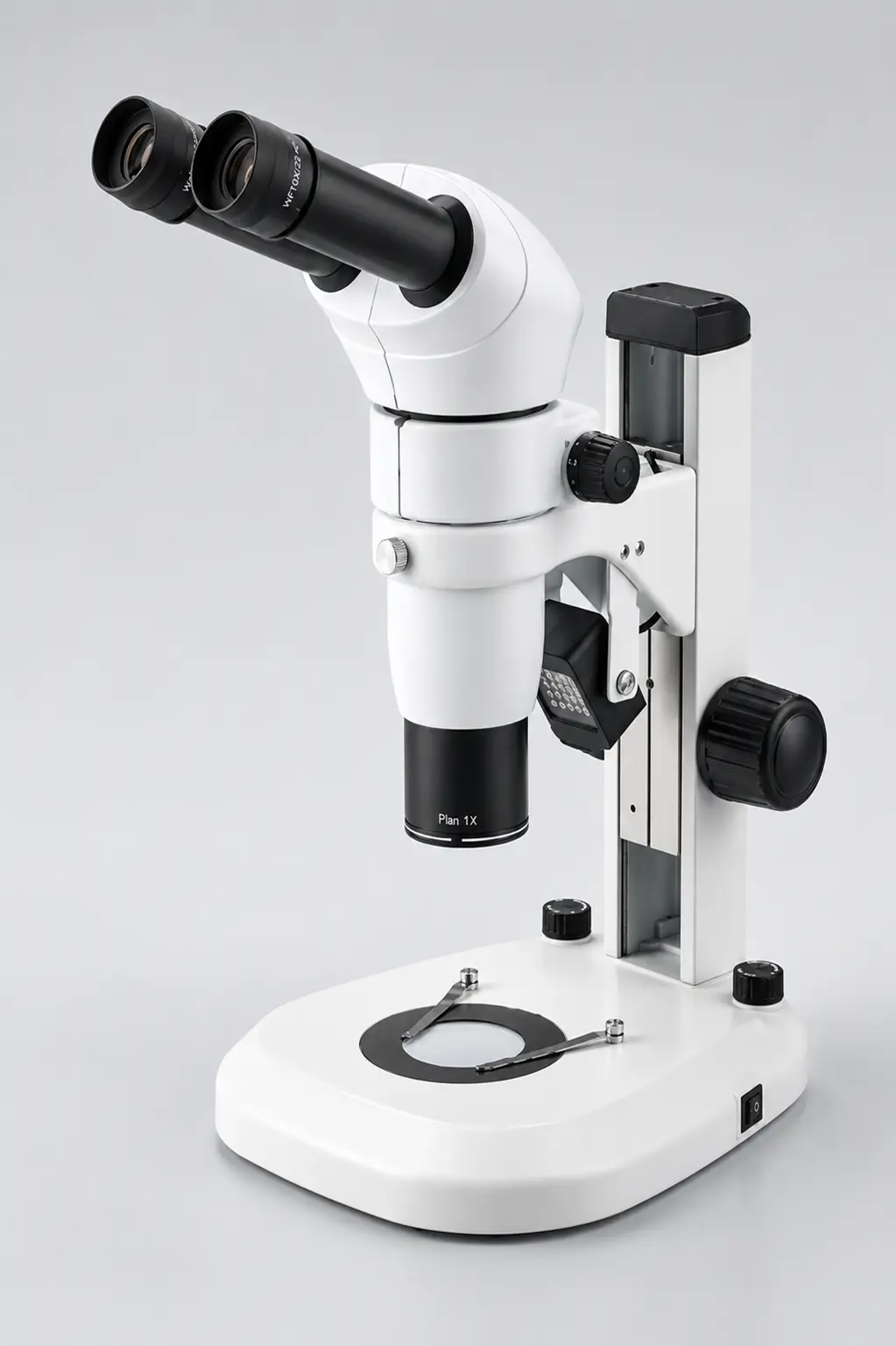
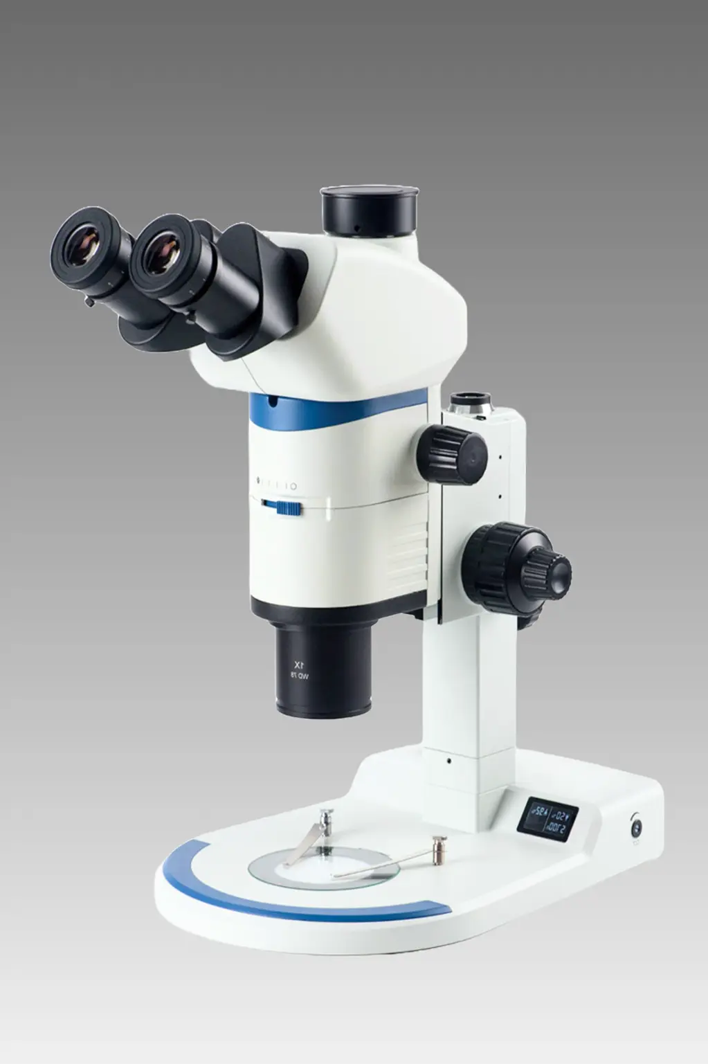
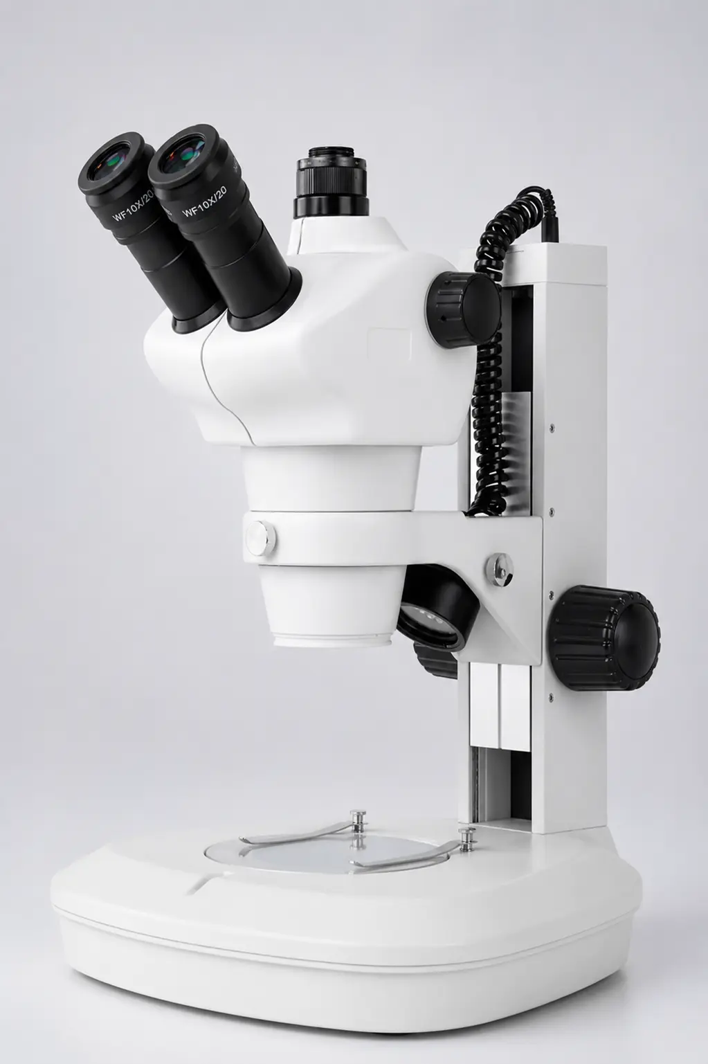
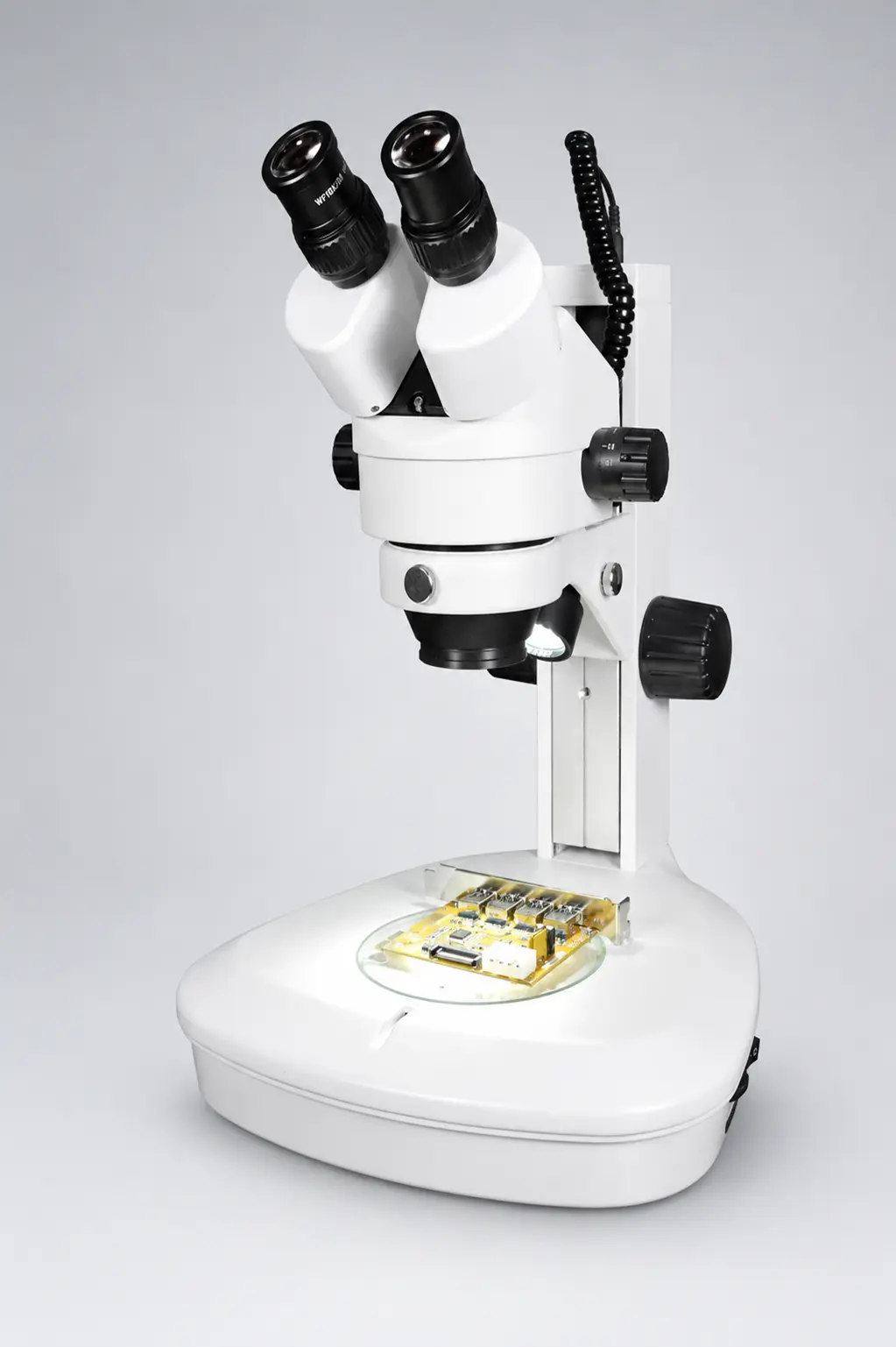
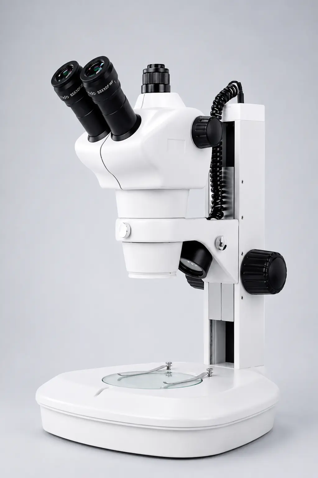
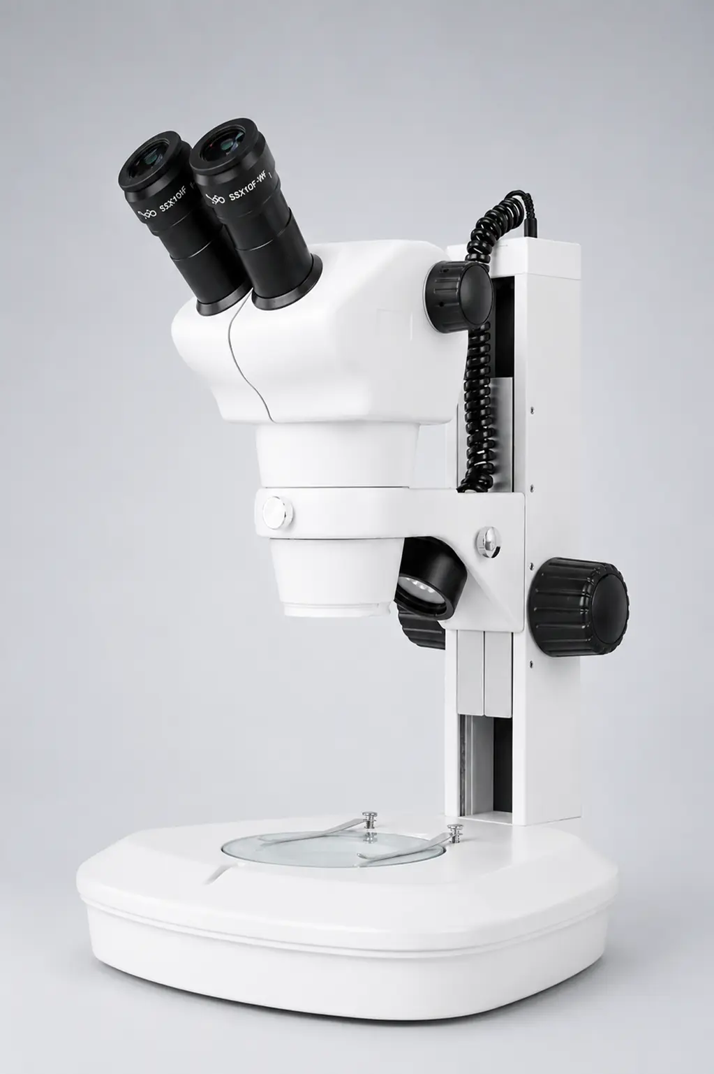
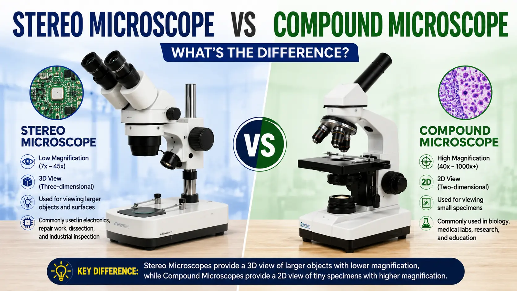
- Stereo zoom microscopes (dissection, electronics, inspection)
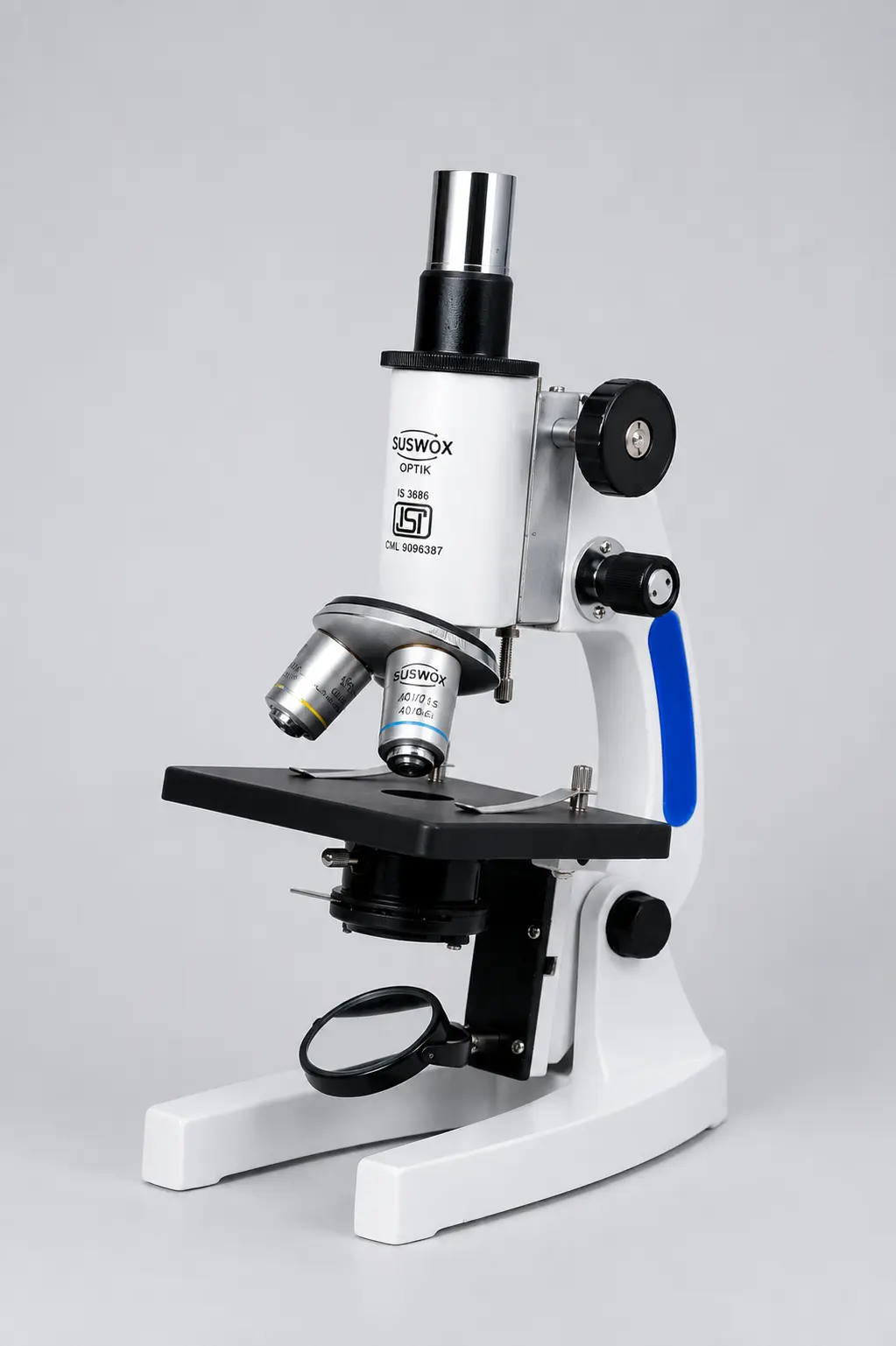
- Student microscopes (school and college labs)
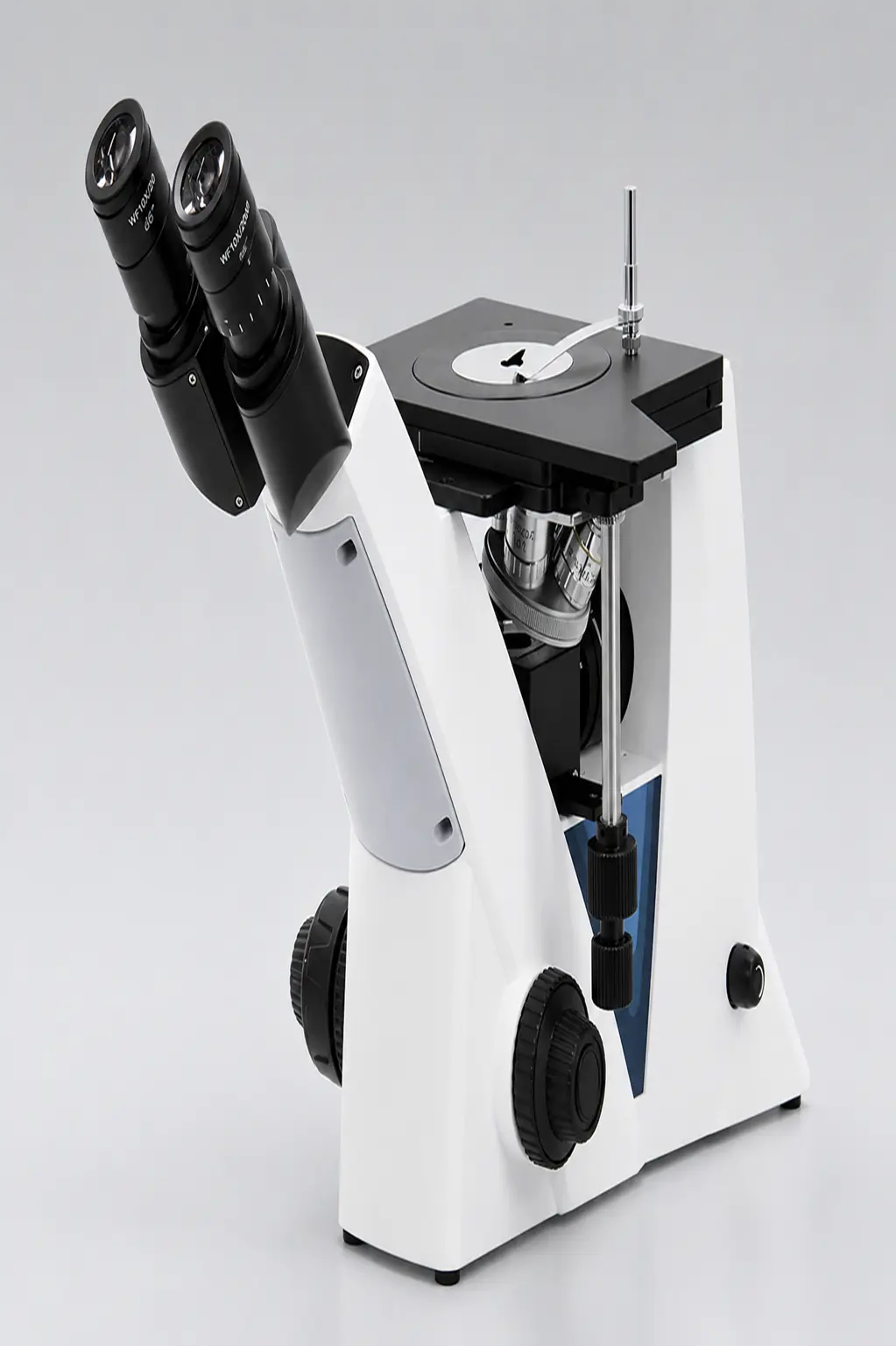
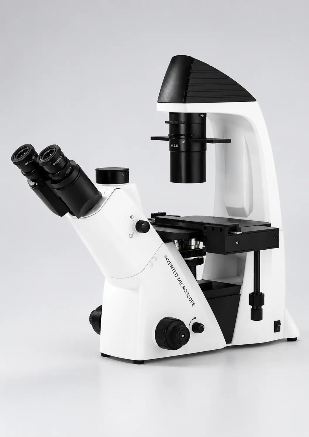
- Inverted microscopes (tissue culture, IVF)
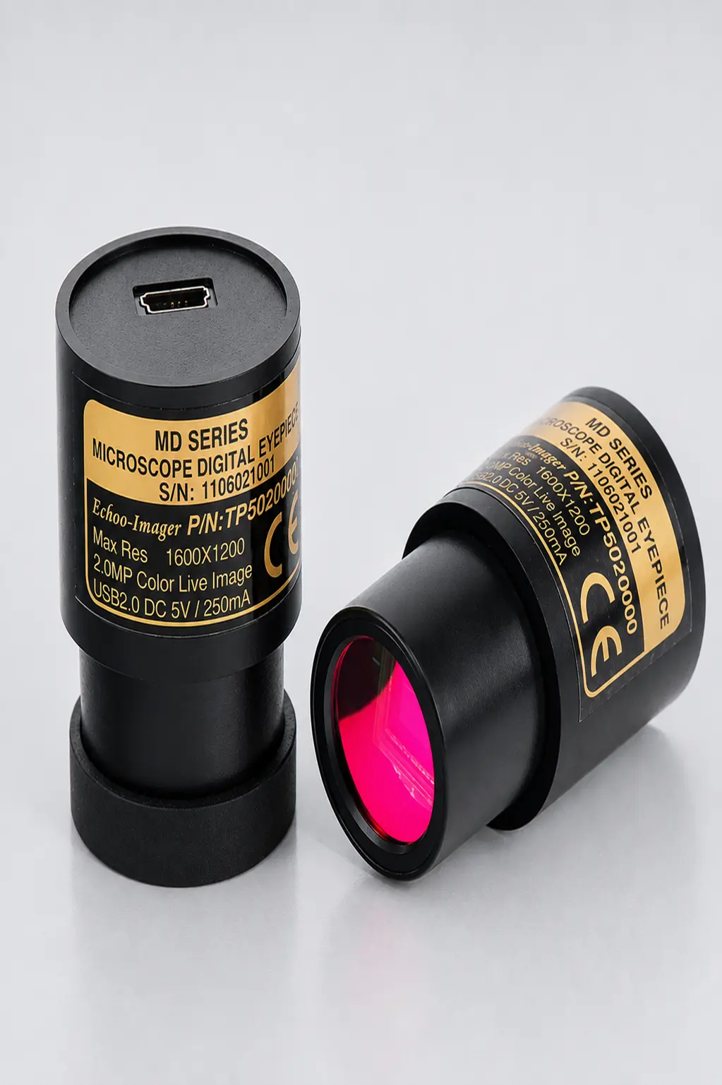
- Digital microscopes (on-screen viewing and documentation)
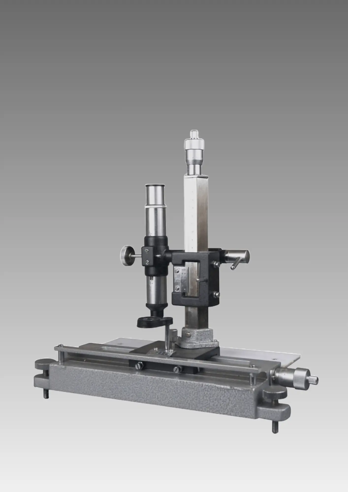
- Travelling microscopes (physics measurement)
- Fluorescence and polarizing microscopes
Main strengths:
- Works on living, moving specimens
- Shows real colour, which matters for stained slides
- Simple to set up and use
- Affordable enough for every school and clinic
- Needs no special room, power or vacuum
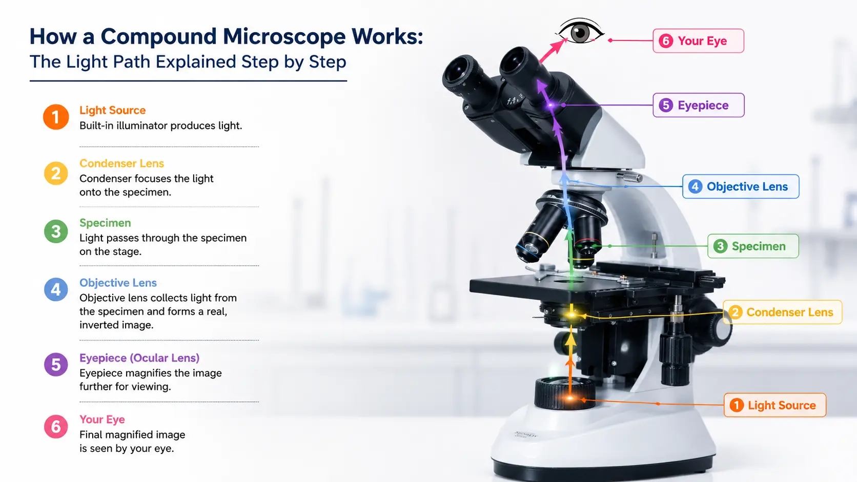
Our guide to the 12 types of microscopes covers each of these categories in detail, and the compound microscope guide explains how the two-lens system actually works.
What is an Electron Microscope?
An electron microscope fires a focused beam of electrons at a sample instead of light. Because electrons have a far shorter wavelength than visible light, they can resolve detail thousands of times finer.
Ernst Ruska built the first one in 1931 in Germany. He received the Nobel Prize in Physics for it — in 1986, fifty-five years later.
There are two main types, and they answer different questions:
| Feature | SEM (Scanning) | TEM (Transmission) |
| How it works | Beam scans across the surface | Beam passes through the sample |
| What you see | 3D surface texture | Flat internal structure |
| Magnification | Up to about 1,00,000x | Up to about 10,00,000x |
| Sample prep | Dried, gold or carbon coated | Ultra-thin sections under 100 nm |
| Typical use | Fracture surfaces, powders, pollen | Viruses, cell organelles, crystal lattices |
The 6 Differences That Actually Matter
1. Why light hits a wall at 200 nanometres
Visible light has a wavelength between roughly 400 and 700 nanometres. You cannot resolve two objects that are closer together than about half that distance — a physical rule known as the Abbe diffraction limit.
So around 200 nm, an optical microscope simply stops separating detail. Push the magnification higher and the picture gets bigger but no clearer. That is called empty magnification, and it is why a listing advertising “2000x” on a school microscope is selling a number, not performance.
Electrons at typical operating voltages have a wavelength thousands of times shorter. The wall moves from 200 nm to around 0.1 nm.
2. Living vs dead samples
An optical microscope can watch a paramecium swim across a drop of pond water. Blood cells move. Cells divide in real time.
An electron microscope cannot do any of that. The sample sits in a vacuum, which means it must be completely dry, and it is usually coated with a thin metal layer as well. Nothing survives that.
So if you need to observe a living process, the choice is made for you regardless of budget.
3. Colour
Optical microscopes show real colour, which is not a cosmetic detail. Gram staining separates bacteria into pink and purple. H&E staining turns nuclei blue and cytoplasm pink. Pathologists read those colours to make a diagnosis.
Electron microscope images are always greyscale, because electrons carry no colour information. The colourful SEM images you see in magazines are coloured afterwards on a computer.
4. Sample preparation
- Optical: place the specimen on a slide, add a cover slip, stain if needed. Minutes.
- Electron: dehydrate, fix, embed, section at under 100 nm with a diamond knife, mount on a grid, coat with gold. Hours to days, with specialised equipment.
That preparation time is a real cost, and it also means the sample you finally view is no longer in its natural state.
5. Infrastructure and running cost
An optical microscope needs a table and a plug point.
An electron microscope needs a dedicated room with vibration isolation, stable temperature, a clean power supply, a vacuum pump system, and a trained operator. Annual maintenance contracts alone often exceed the entire cost of a good optical microscope.
6. Who actually uses which
| Setting | Instrument Used |
| School and college labs | Optical |
| Pathology and diagnostic labs | Optical |
| Hospitals and clinics | Optical |
| Water and food testing | Optical |
| Industrial QC and electronics | Optical (stereo) |
| Nanotechnology research | Electron |
| Virology and vaccine research | Electron |
| Semiconductor failure analysis | Electron |
| Materials and metallurgy research | Both |
Look at that list honestly and one thing becomes clear. Optical microscopes carry almost the entire daily workload of science in India. Electron microscopes handle a narrow band of research questions that nothing else can answer.
Which One Do You Actually Need?
Choose an optical microscope if you:
- Run a school, college, hospital, pathology or testing lab
- Need to view cells, bacteria, tissue, blood or water samples
- Want to observe living organisms
- Have a normal room and a normal budget
- Need results the same day, with minimal preparation
You need an electron microscope if you:
- Study viruses, nanoparticles or atomic structure
- Do materials research, semiconductor analysis or nanotechnology
- Require resolution below 200 nanometres
And here is the practical reality for the second group: almost nobody buys one. Most Indian researchers book time at a shared facility instead — the Sophisticated Analytical Instrument Facilities set up under the Department of Science and Technology, or the central instrumentation centres at IITs, NITs, CSIR labs and larger state universities. You send the sample, pay per hour, and get images back.
Which is far more sensible than a ₹2 crore instrument sitting idle for eleven months a year.
Frequently Asked Questions
Which microscope has the highest magnification?
The transmission electron microscope (TEM) has the highest magnification, reaching roughly 10,00,000x with resolution near 0.1 nanometre. Among optical microscopes, the compound microscope goes highest at about 1000x to 1500x using a 100x oil immersion objective.
Can an optical microscope see viruses?
No. Most viruses measure between 20 and 300 nanometres, which is at or below the 200 nm resolution limit of visible light. Viruses are imaged using transmission electron microscopes. Bacteria, being much larger, are clearly visible at 1000x on a standard compound microscope.
Is an electron microscope better than an optical microscope?
Neither is better — they solve different problems. An electron microscope gives far higher resolution but cannot show living or coloured samples and costs a hundred times more. For routine diagnostics, teaching and inspection, an optical microscope is not just adequate, it is the correct instrument.
Why can’t electron microscopes show colour?
Colour is a property of visible light, and electron microscopes do not use light. The detector records electron intensity, which produces a greyscale image. Colour seen in published SEM images is added digitally afterwards.
What is the price of an electron microscope in India?
Scanning electron microscopes generally start around ₹40 lakh and rise well past ₹1 crore depending on configuration, while transmission electron microscopes typically run into several crores. Installation, the vacuum system and annual maintenance are additional.
Has the 200 nm limit ever been broken by light microscopes?
Yes, partly. The 2014 Nobel Prize in Chemistry went to Eric Betzig, Stefan Hell and William Moerner for super-resolved fluorescence microscopy, which uses fluorescent tricks to push light microscopy below the diffraction limit. Those systems are research-grade and priced accordingly.
Buying an Optical Microscope in India
For the vast majority of laboratories, the practical question is not optical versus electron. It is which optical microscope, and from where.
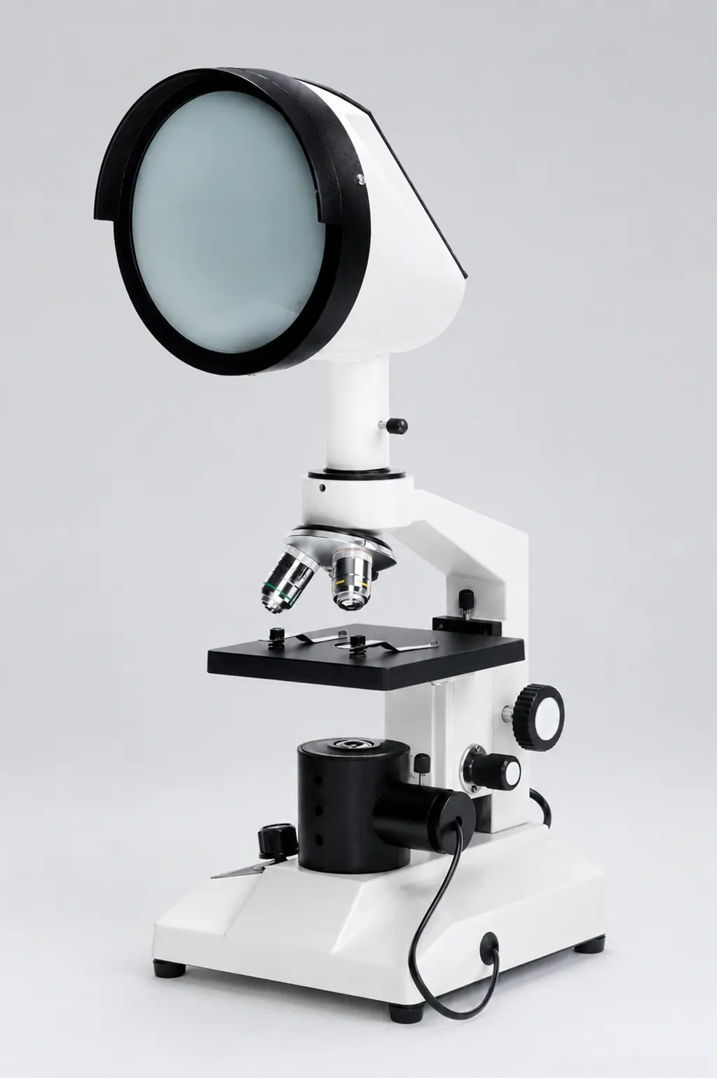
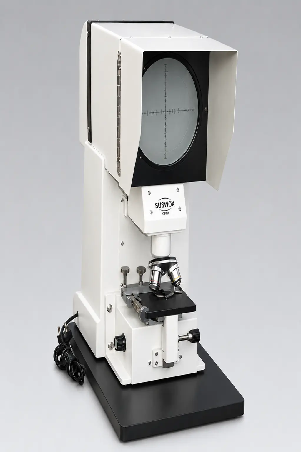
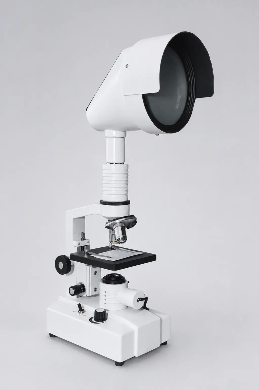
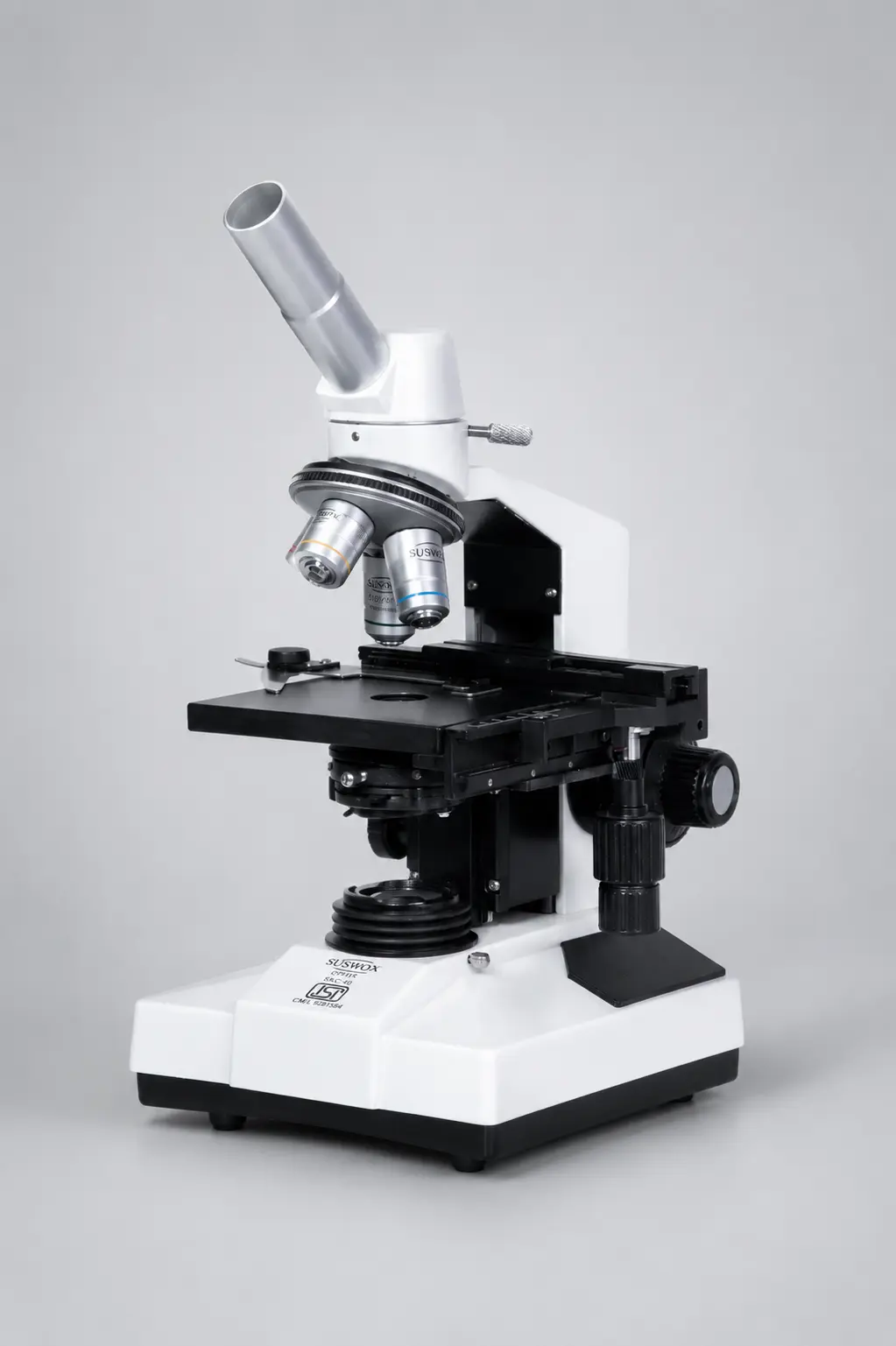
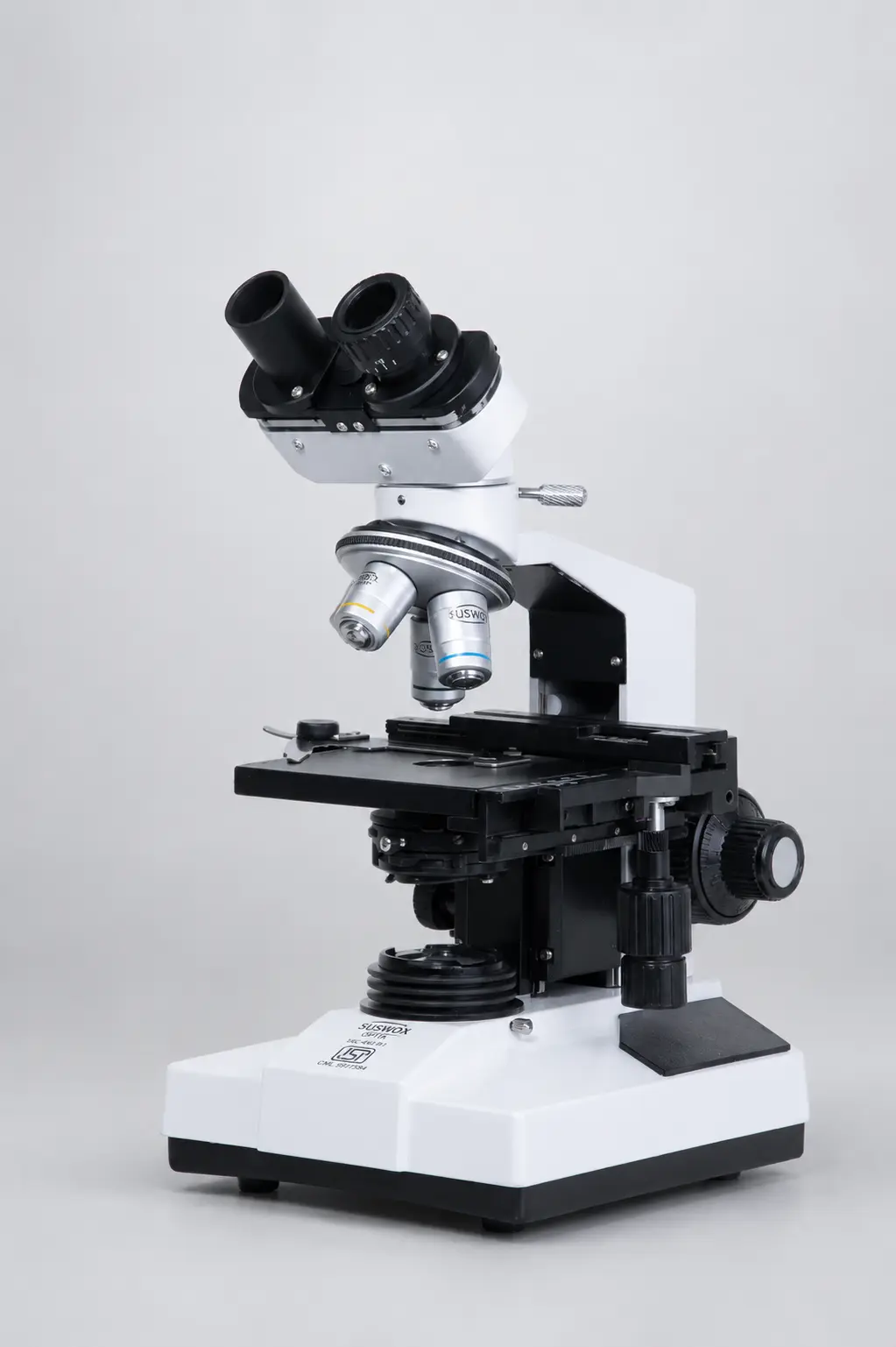
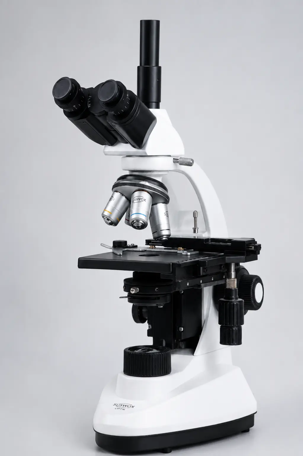
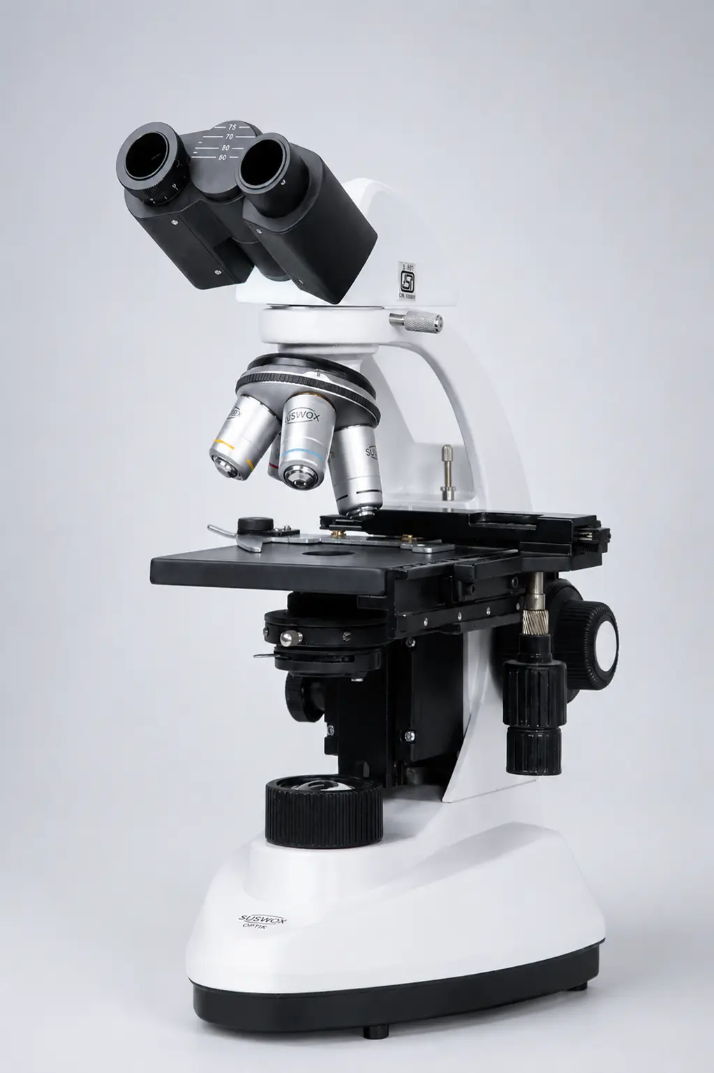
Suswox (Sudheer Scientific Works) is a microscope manufacturer in India based at 1265 Bengali Mohalla, Ambala, Haryana, manufacturing optical microscopes since 1973. Ambala has been the country’s scientific instruments hub for decades. The range covers compound laboratory microscopes in the SC-300, SKC and CXL series, student microscopes in the SK series, stereo zoom, inverted, digital, travelling, projection and operating microscopes.
Suswox instruments are in daily use at AIIMS Bathinda, AIIMS Nagpur, AIIMS Bilaspur, BHU, Kurukshetra University, MDU Rohtak and the Haffkine Institute.
What buying direct gets you, compared to trading-only microscope suppliers in India:
- Factory pricing with no reseller margin
- Spare parts and service for the working life of the instrument
- Configuration to your requirement — objectives, illumination, head type, stage
- Anti-fungal treated optics, which matter in humid Indian conditions
- Matched optics across every unit in a bulk institutional order
- GeM listing and tender documentation support
That last point is where established microscope manufacturers differ from resellers. A university ordering fifteen identical microscopes with one service contract needs a factory behind it, not a stock list.
Suswox — Sudheer Scientific Works 1265 Bengali Mohalla, Ambala (HR), India 133001 +91 8727015757 | +91 9416025615 | sales@suswox.com
Browse all microscopes · Compare stereo vs compound · Request a quote
The Short Answer
If your sample is alive, coloured, or larger than about 200 nanometres, an optical microscope is the right tool — and the only one you are likely to own.
If it is smaller than that, no amount of magnification will help. You need electrons, and you probably need to book time at a shared facility rather than buy the instrument.