
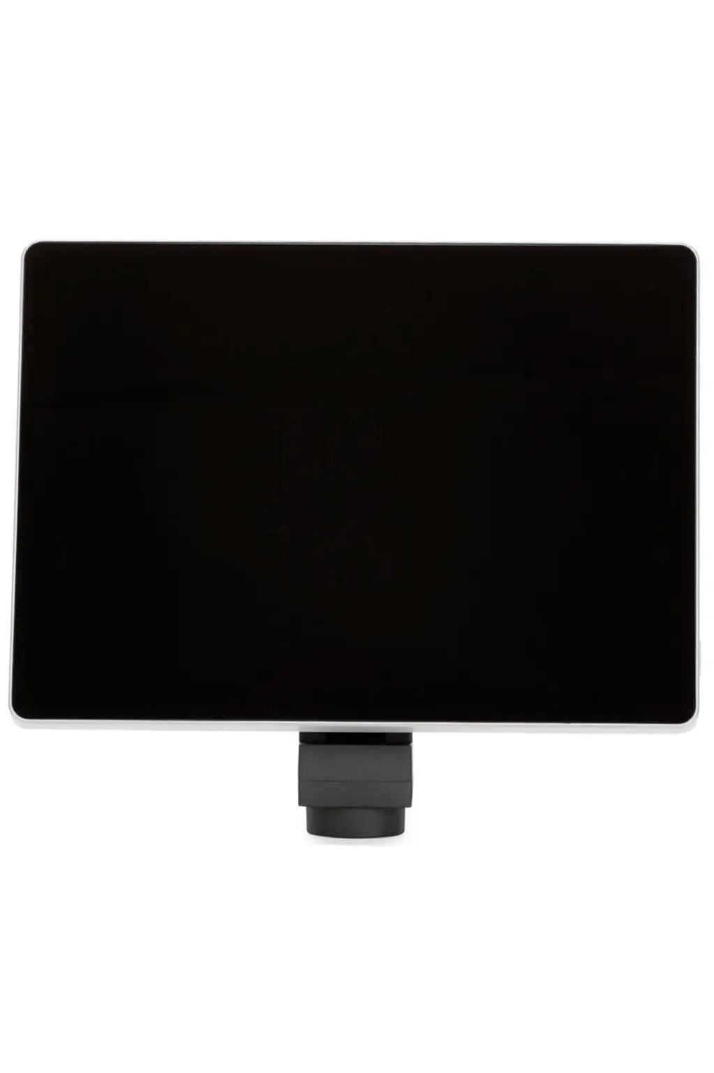
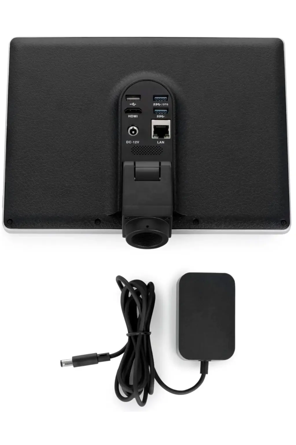
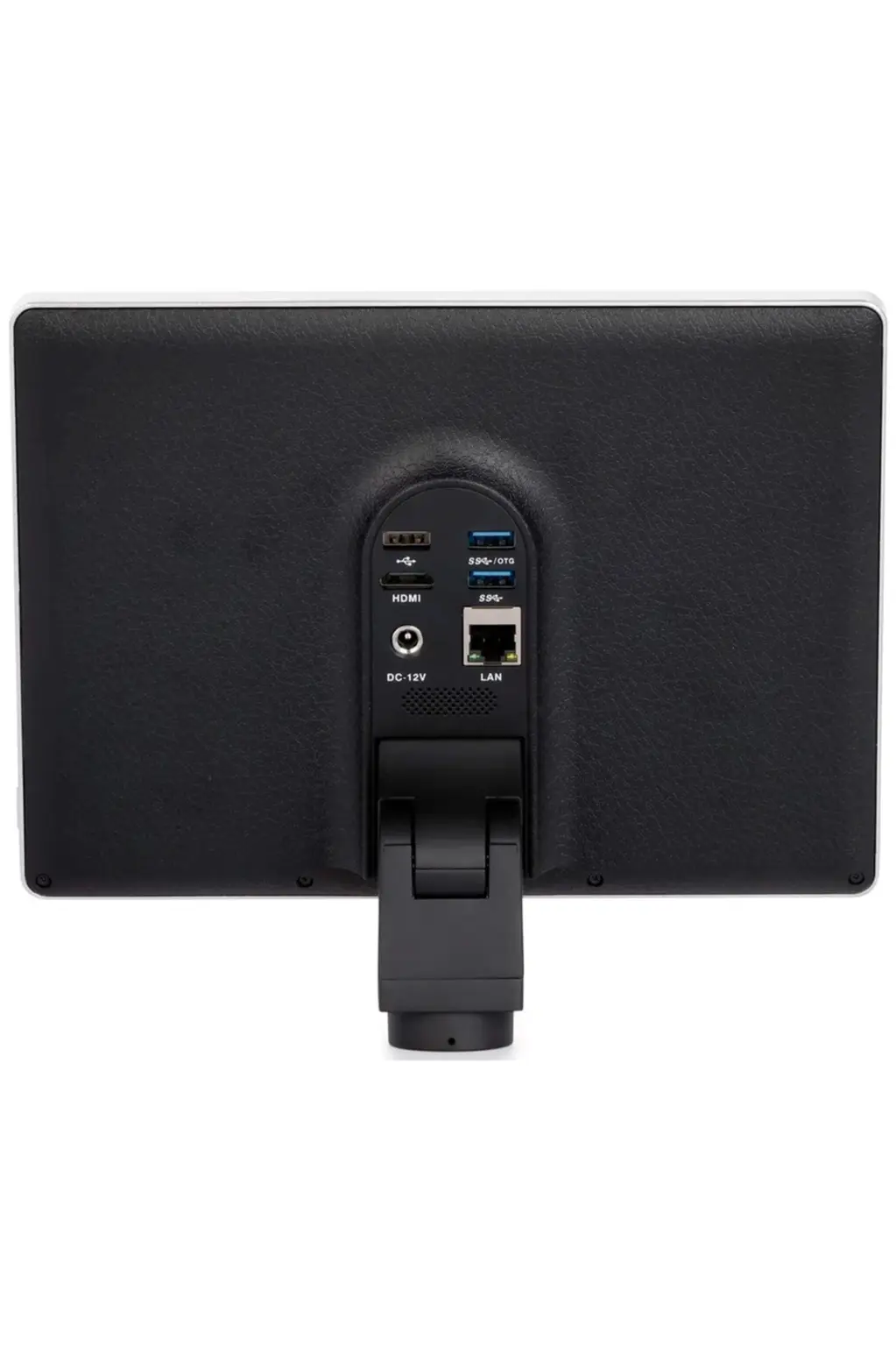
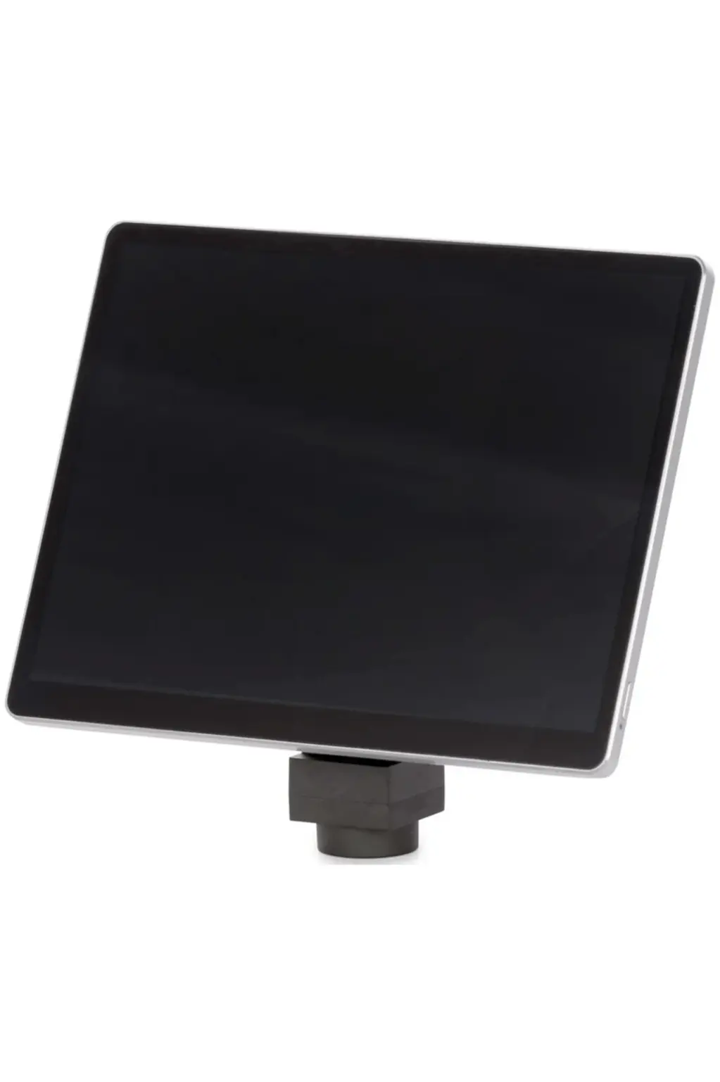
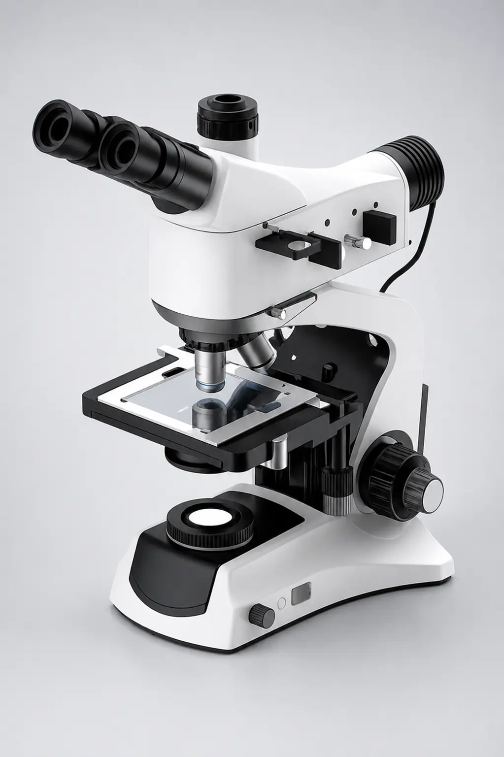
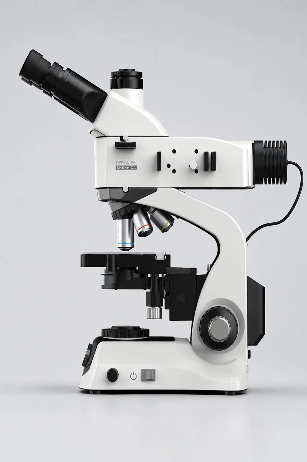
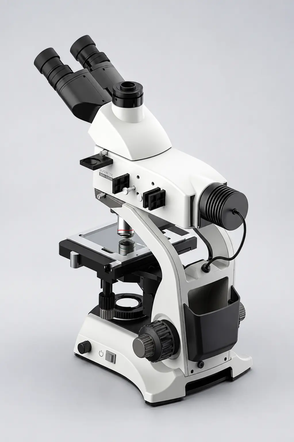
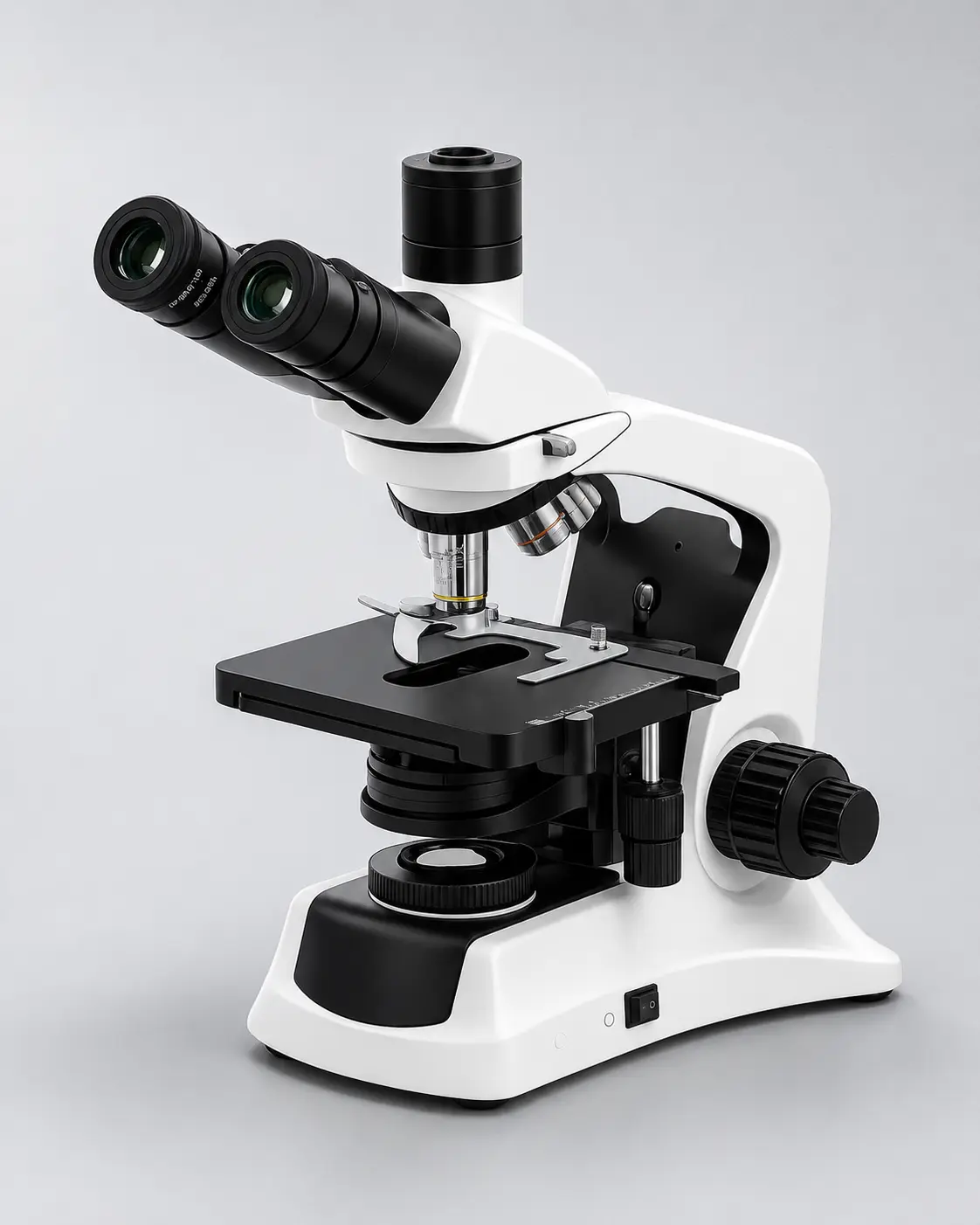
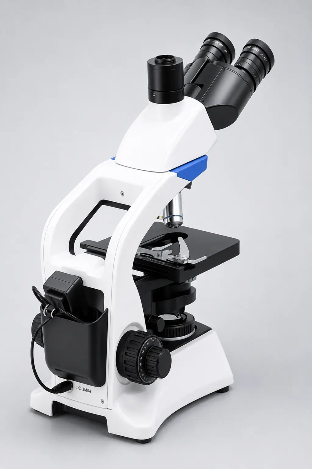
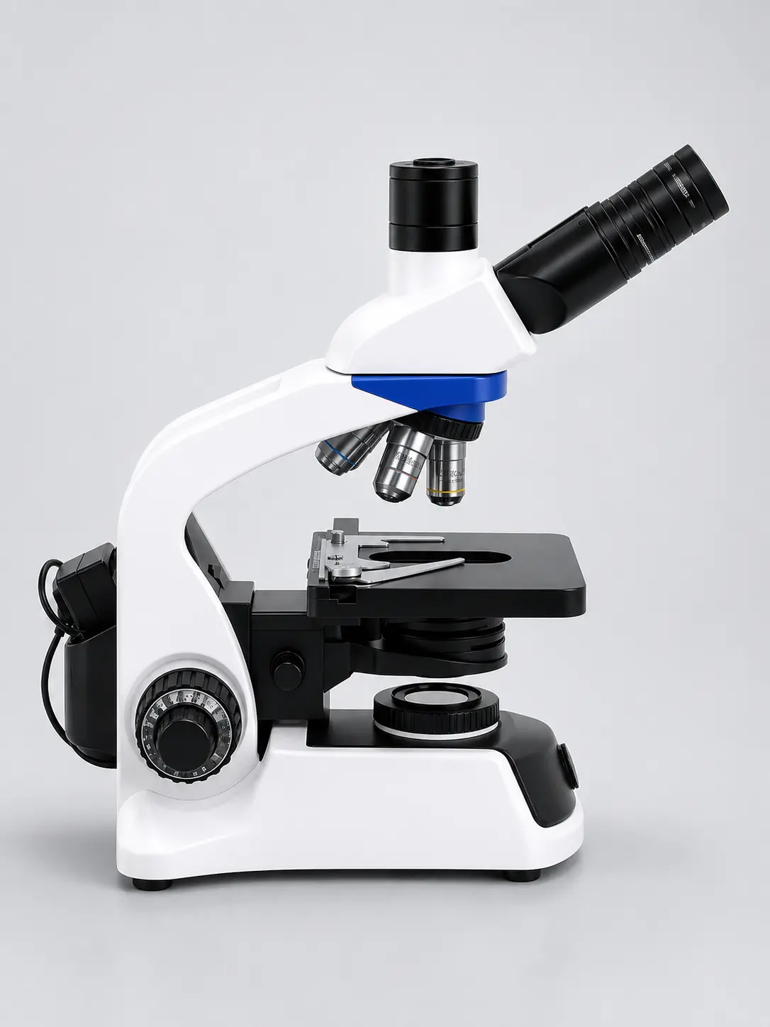
AT Series Specification
Maximum total magnification
ABBE double-lens condenser
Rated LED lamp life
SMPS, handles Indian supply swings
| Specification | Details |
|---|---|
| OS | Android 11 |
| CPU | RK3568 |
| Memory | 2GB |
| Internal Storage | 32GB |
| External Storage | USB |
| Display Type | Touchscreen LCD |
| Screen Size (diagonal) | 10.5″ |
| Display Resolution | 1920 x 1280 |
| Display Mount | 360° rotation, 90° tilt |
| Sensor Type | CMOS |
| Sensor Optical Format | 1/2″ |
| Active Pixels | 8M (16:9) |
| Pixel Size | 2.2µm x 2.2µm |
| Active Sensor Area | 7.74mm x 4.38mm |
| Shutter | Electronic rolling shutter |
| Photo Capture Resolution | 3840 x 2160 |
| Video Capture Resolution and Maximum Framerate | 30fps @3840 x 2160 |
| Connectivity | HDMI, USB 3.0, LAN/WLAN |
| Output Ports | 1x USB Mouse port, 2x USB port, 1x HDMI Port, 1x LAN Port |
| Lens-mount | C-mount |
| Power | 12V/1A, 110–220VAC |
Digital microscopes combine live imaging and documentation: enabling shared observation, streamlined workflows, and precise inspection across industries and education.
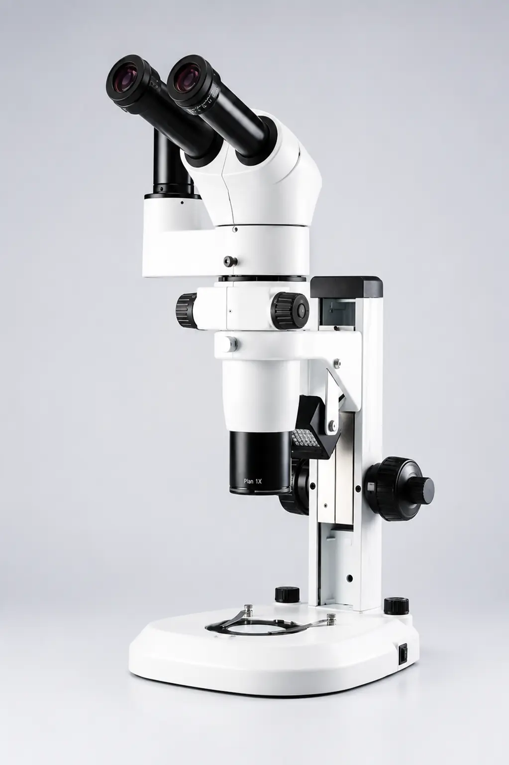
Ideal for detailed inspection of components, assemblies, samples and manufactured products. Live digital imaging enables operators to examine fine details directly on a monitor while supporting efficient quality-control workflows.
Suitable for biological, material and scientific microscopy where high-quality digital images are required. Camera-based systems enable observation, image capture, documentation and further analysis.
Designed for inspection of PCBs, solder joints, connectors, electronic components and miniature assemblies. Digital viewing provides a comfortable working environment for prolonged inspection and precision rework.
Excellent for teaching laboratories, demonstrations and practical training. Multiple users can view the live microscope image simultaneously on a display, making microscopic structures easier to explain and discuss.
Useful for production-line inspection, component verification, surface examination and precision assembly. Digital imaging helps operators identify defects, compare samples and maintain visual inspection records.
Ideal for capturing, storing and sharing microscope images and videos for reports, presentations, research records and quality documentation. Compatible imaging solutions can extend the system for measurement and advanced image analysis.
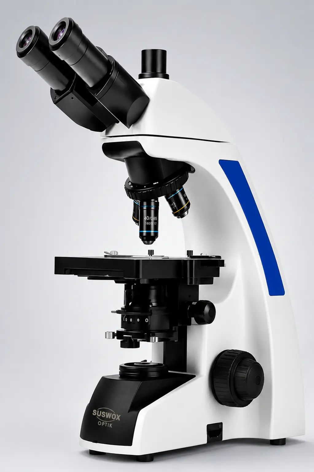
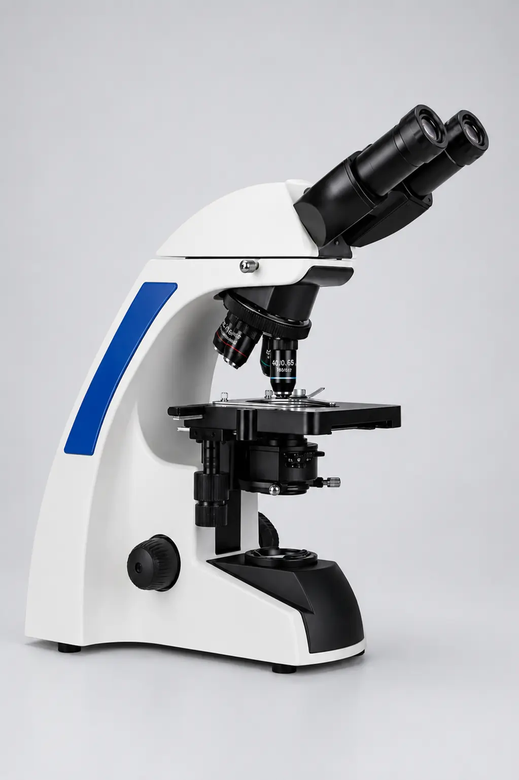
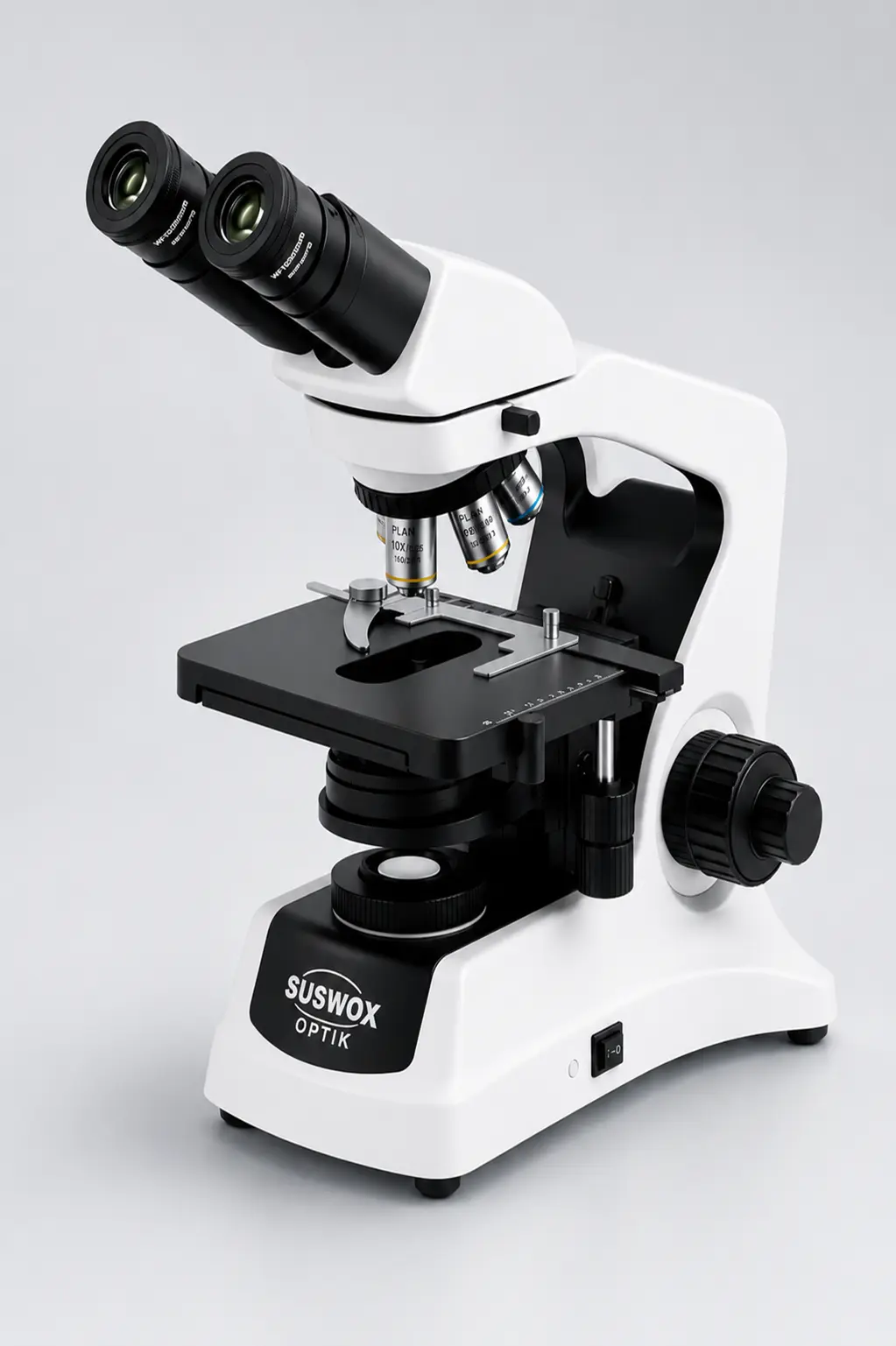
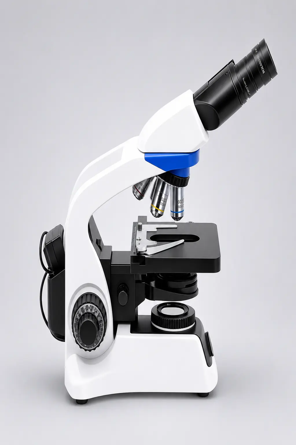
1000×. The WF10X eyepieces multiply with the 100X oil immersion objective, giving 40×, 100×, 400× and 1000× across the four positions on the nosepiece. Beyond 1000× you gain no additional detail with this optical system — you are only enlarging an image the numerical aperture has already limited.
You need it for the 100X objective. Without oil filling the gap between the coverslip and the front lens, light bends away at the glass–air boundary and the effective N.A. drops well below 1.25 — the image goes dim and soft. A vial is included in the box. Wipe the objective with lens tissue after each session.
Semi-plan objectives give a flat, sharp image across roughly 80% of the field, with slight softening at the extreme edge. Plan objectives correct the full field but cost significantly more. For routine screening and teaching, semi-plan is the practical choice; for photomicrography where edge-to-edge sharpness is published, ask us about the plan upgrade.
LED for almost every lab. It runs at 50,000 hours against roughly 1,000 for halogen, stays cool so it does not dry your specimen, and holds colour temperature as you dim it. Halogen remains available if your protocol was validated on it or you need its warmer spectrum for particular stains.
Yes. The sliding binocular head accepts our XCAM Mini and X Cam C-mount cameras through an adapter. If documentation is a daily requirement rather than an occasional one, consider a trinocular model instead so you keep both eyepieces and the camera port live at the same time.
Stock units dispatch in 3–5 business days. We ship across India by surface and air freight, and export with wooden crating on request. Institutional and tender orders with specific documentation requirements are handled by our sales desk — call +91 8727015757.
The unit carries a one-year warranty on body, optics and electricals. We stock spares — objectives, eyepieces, LED assemblies, stage parts — for ten years from the date of manufacture. Most issues are resolved over a video call; parts ship direct to your lab.
Pricing depends on configuration (LED or halogen, objective set, camera adapter) and order quantity, so we quote per enquiry rather than list a single figure. Send your requirement through the form below or on WhatsApp and you will have a written quotation with GST and freight the same working day.
Lets Have A Chat