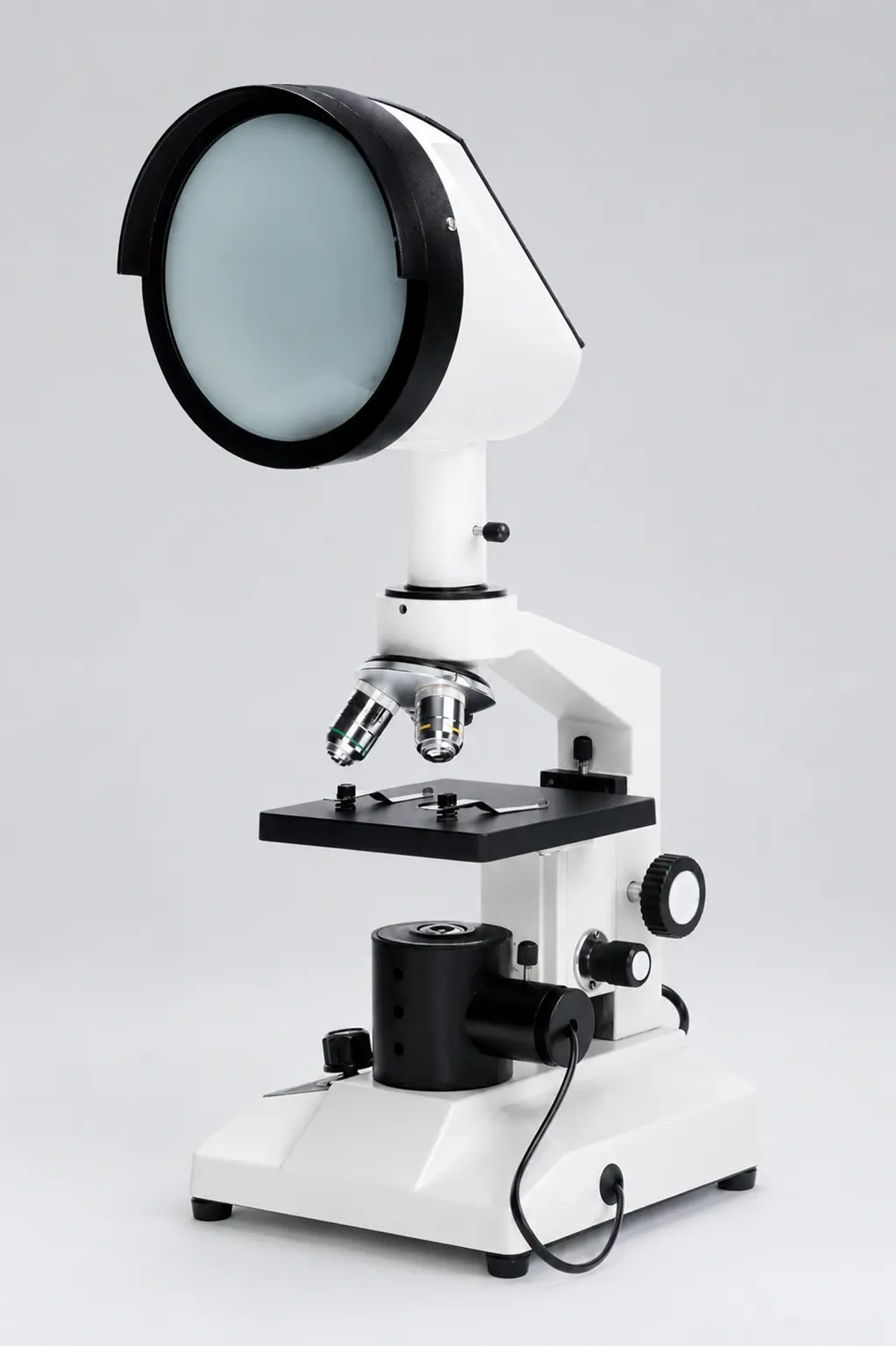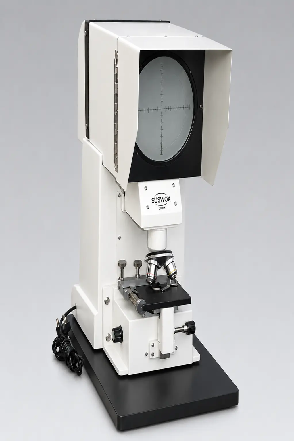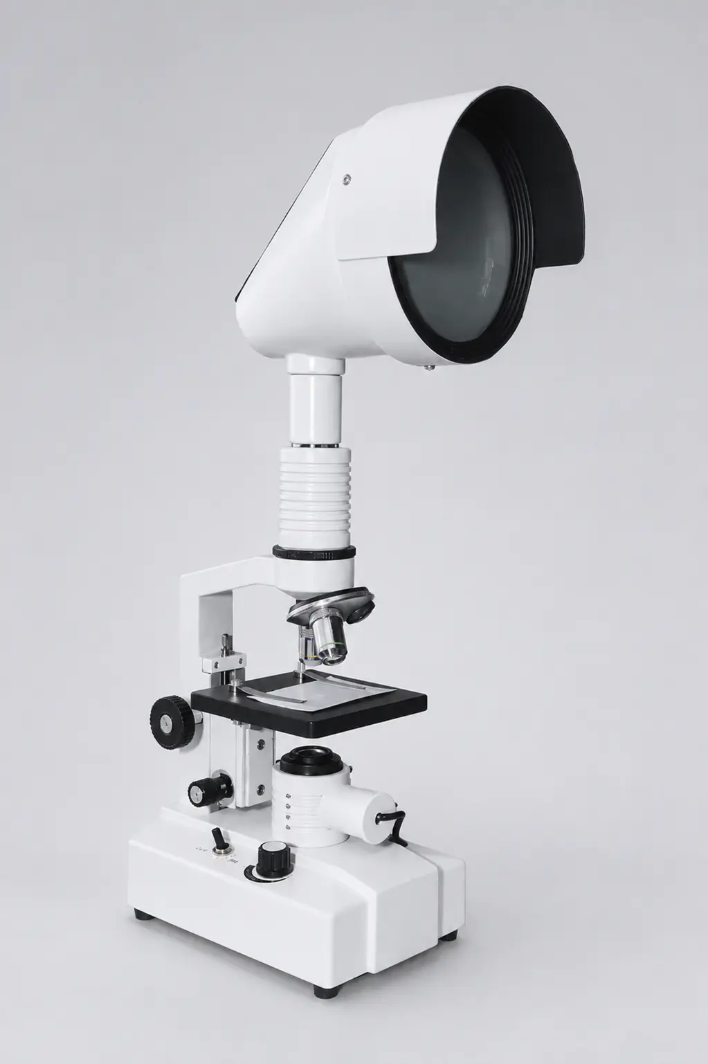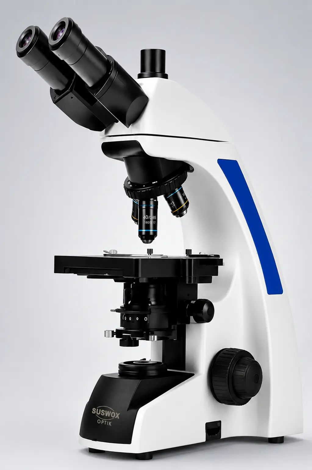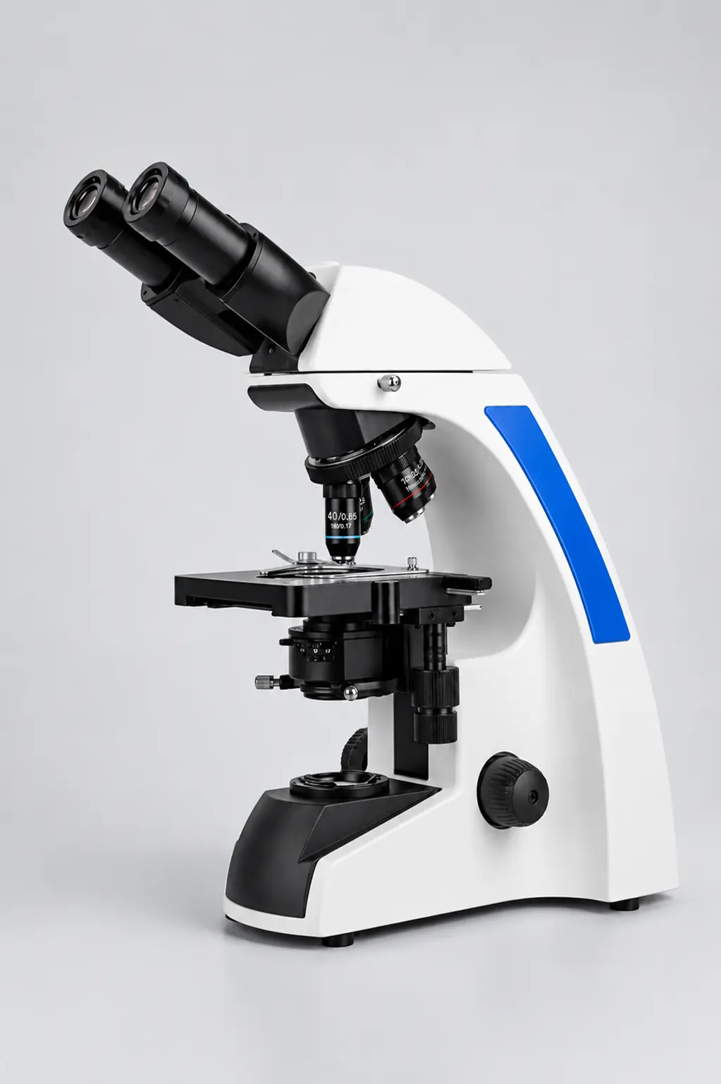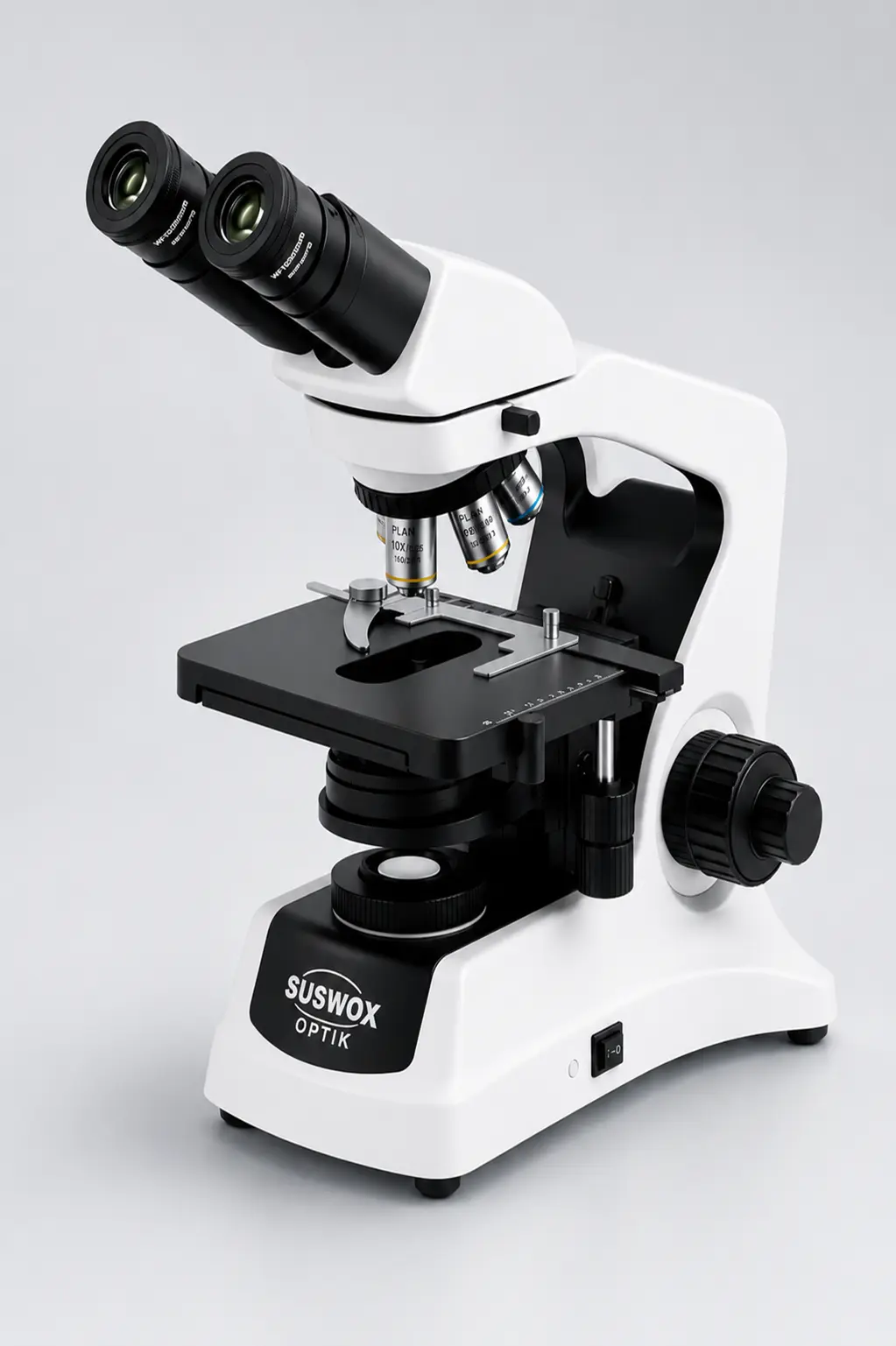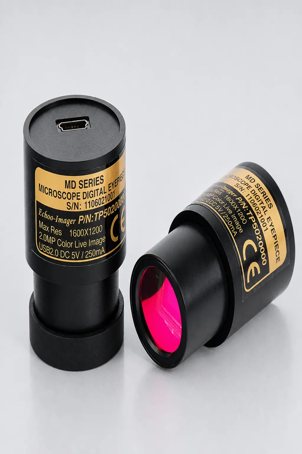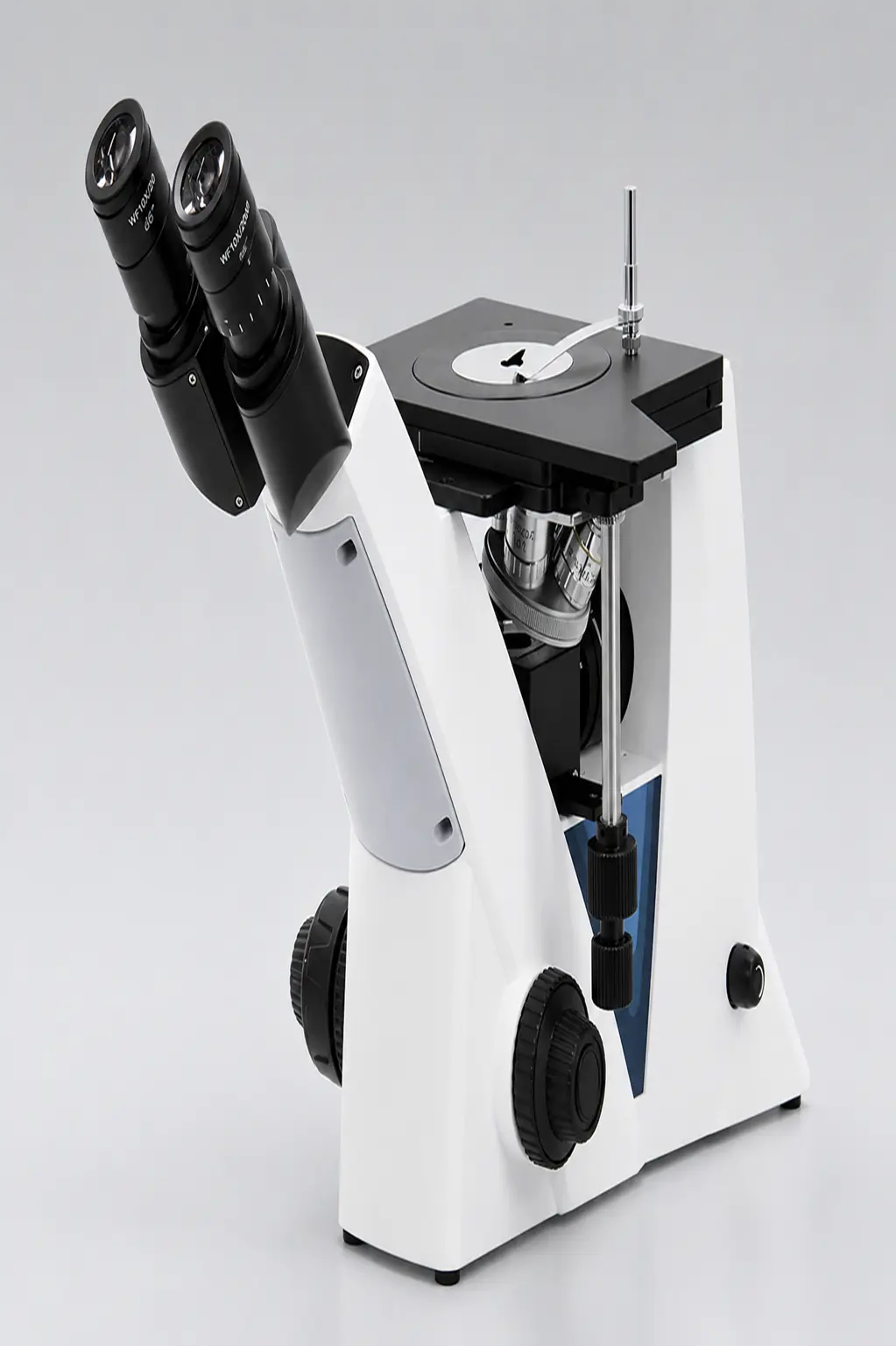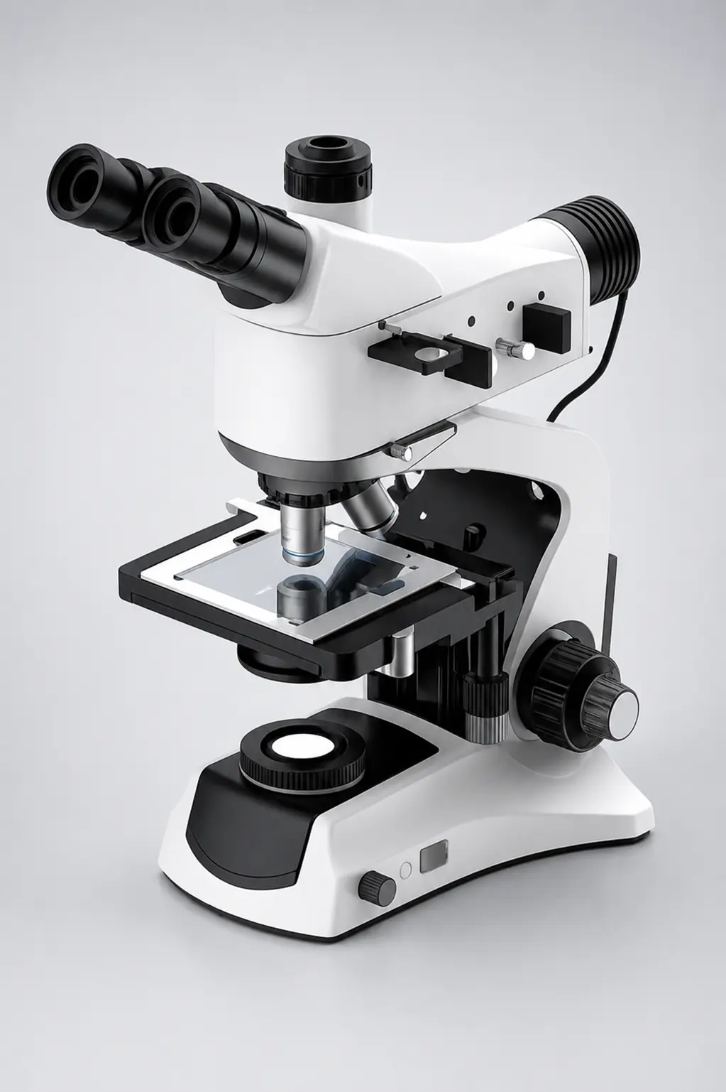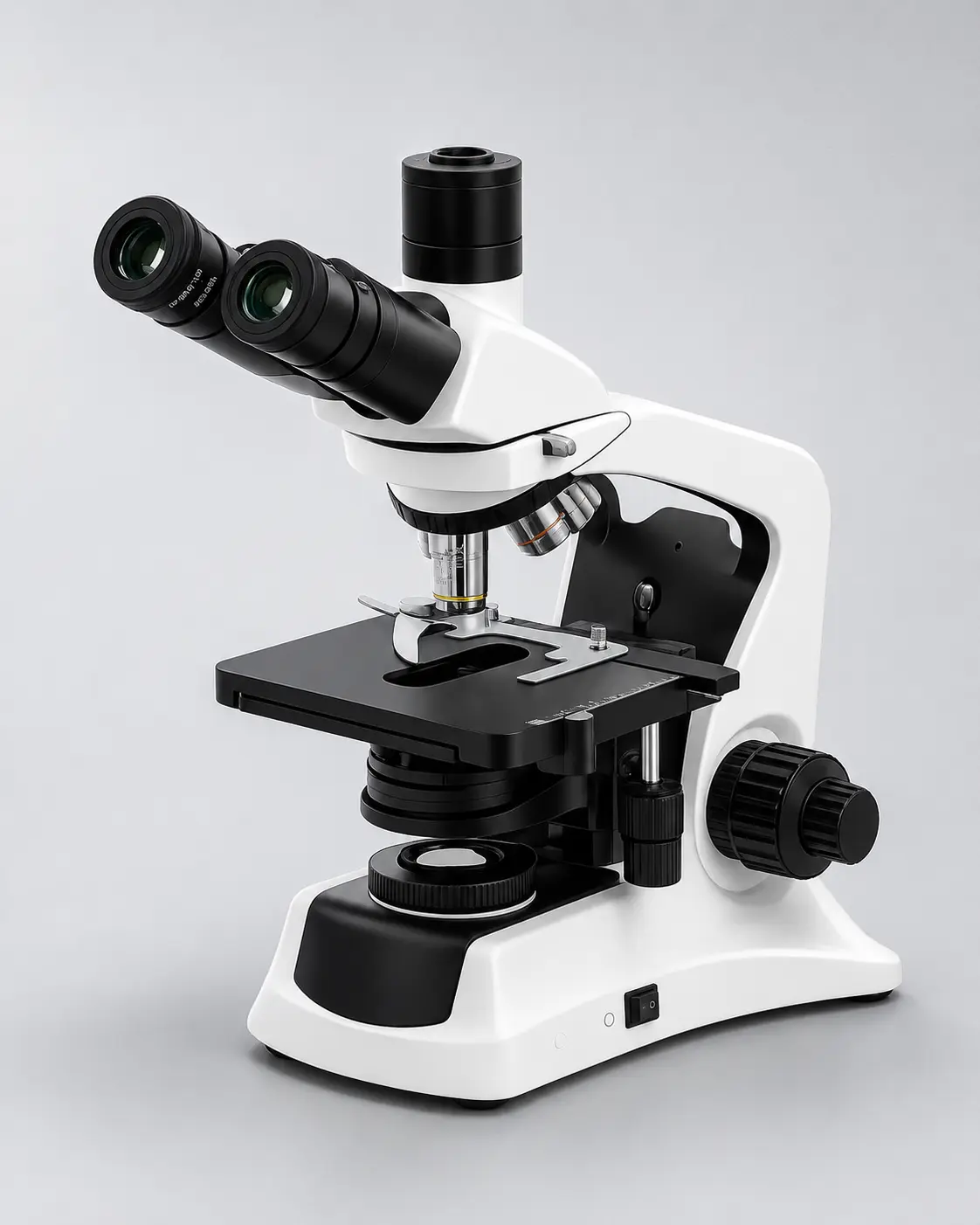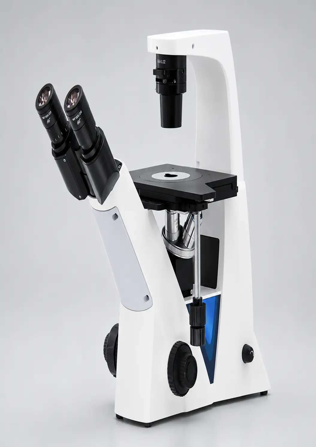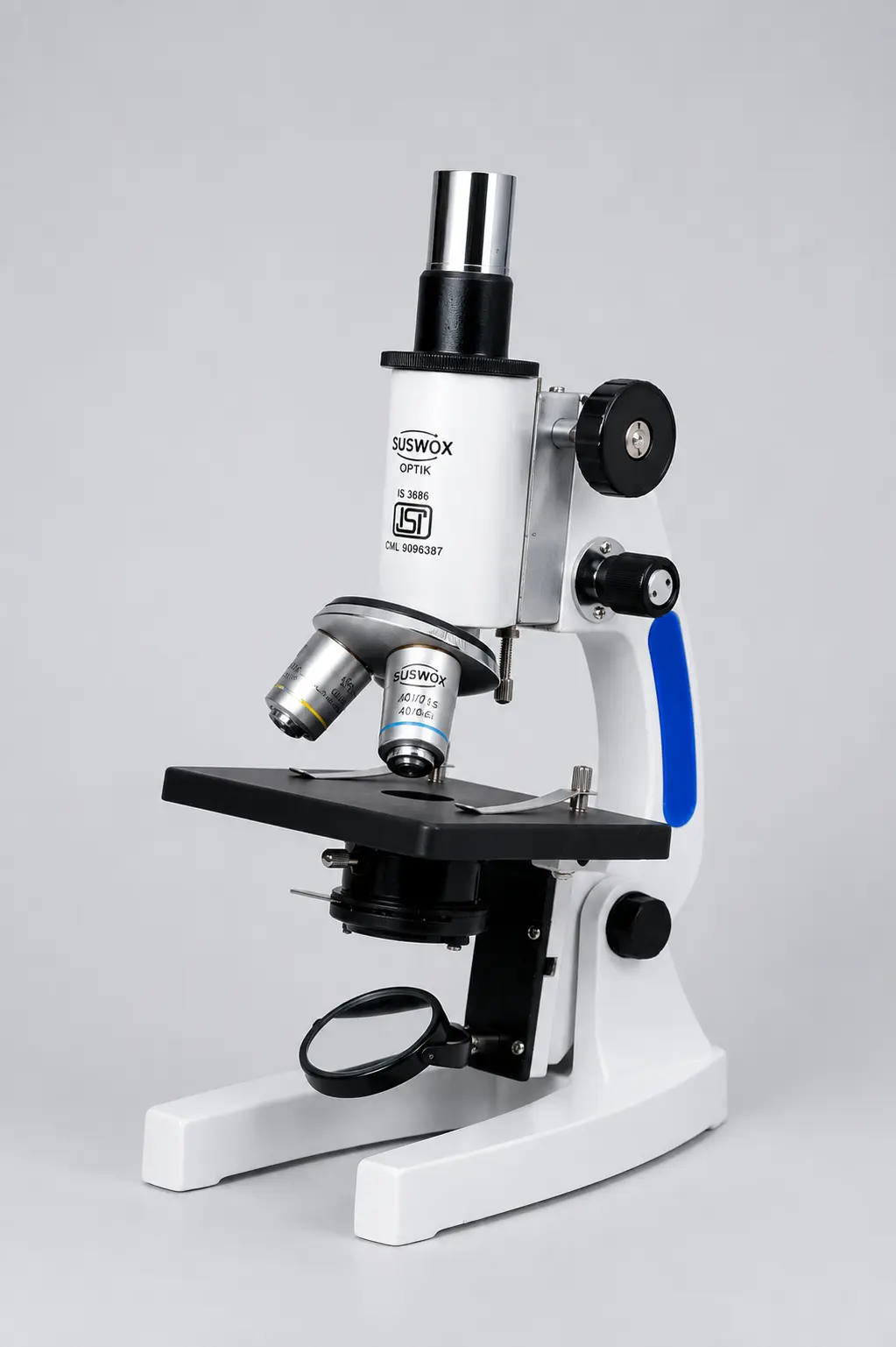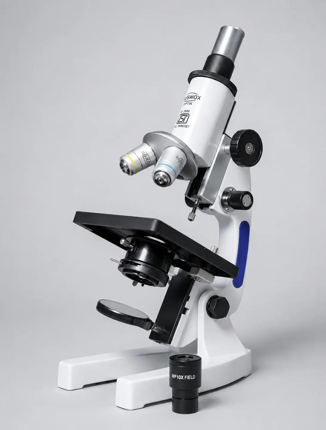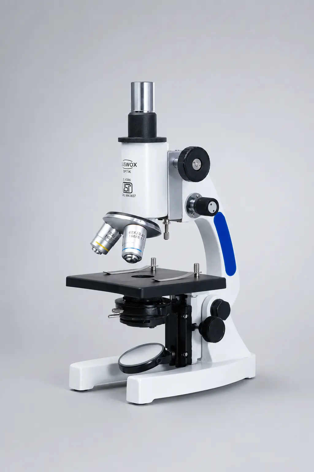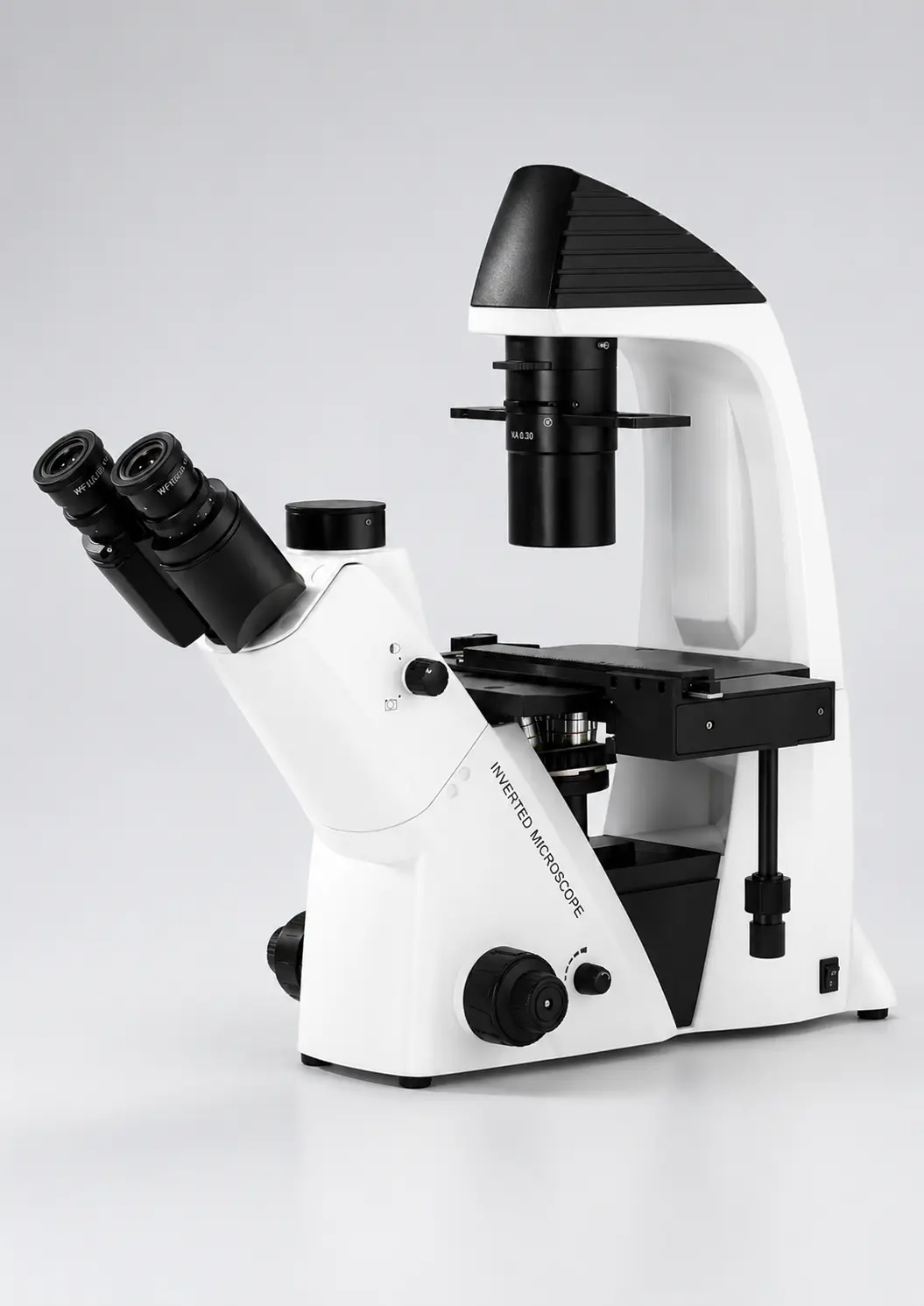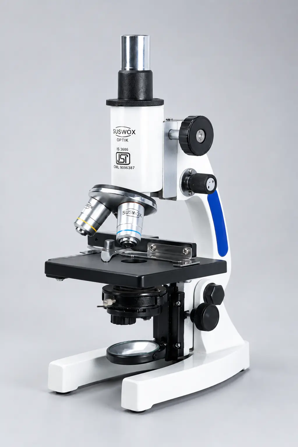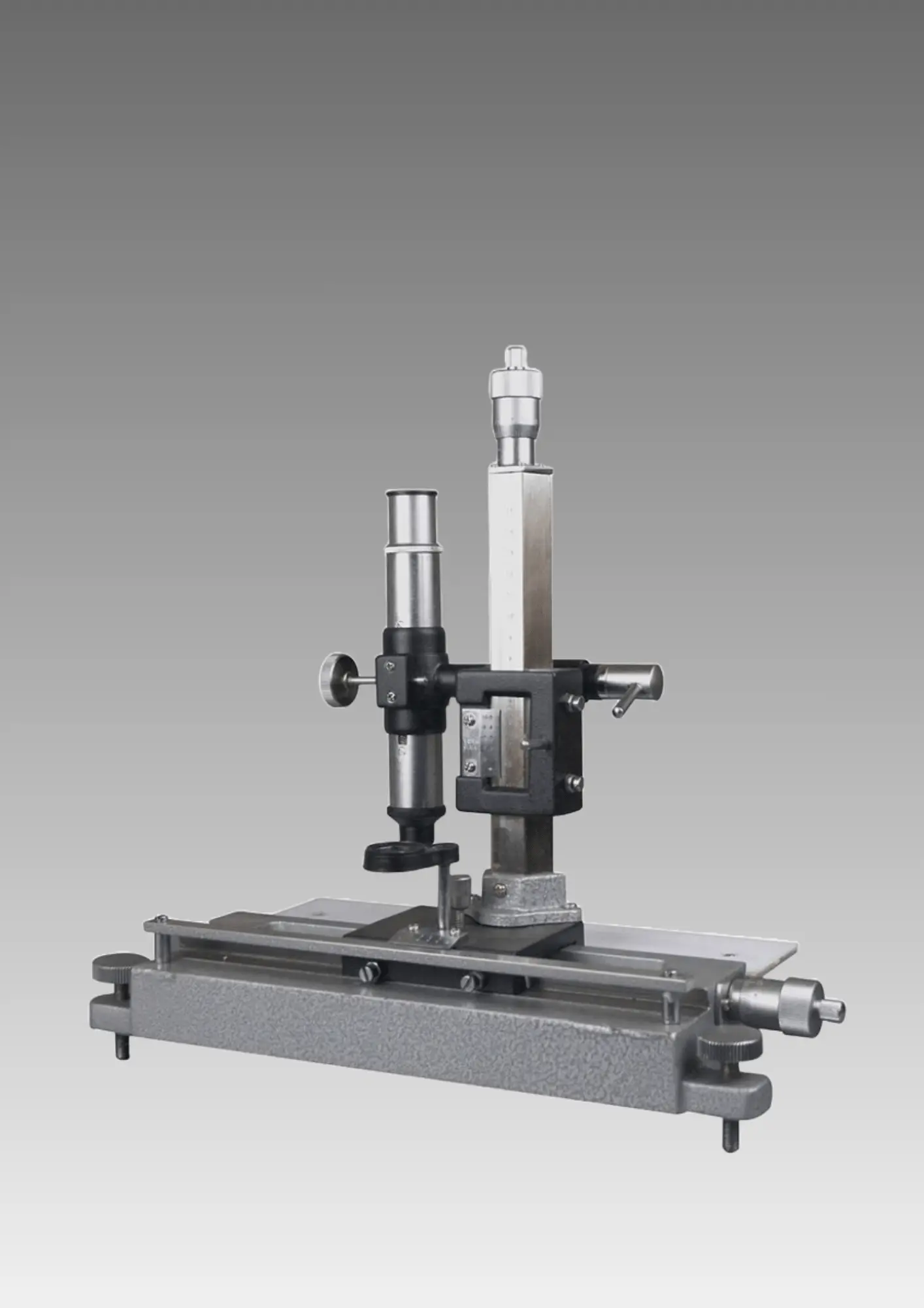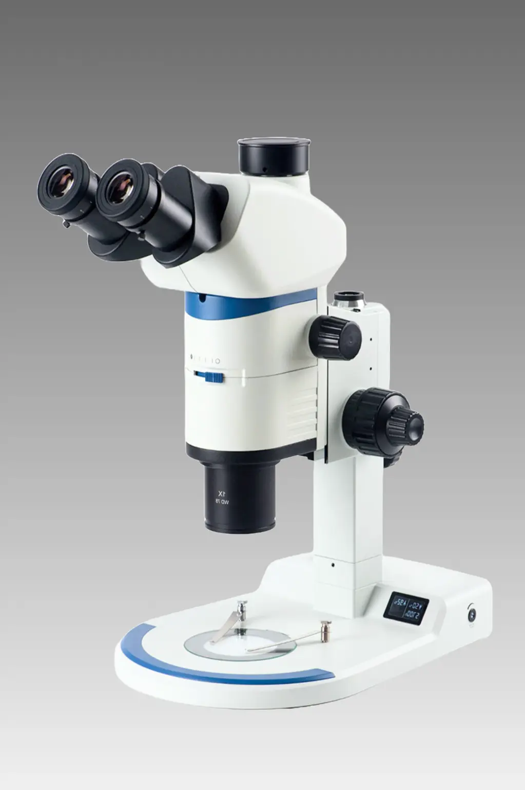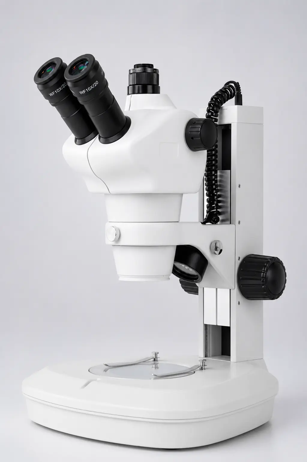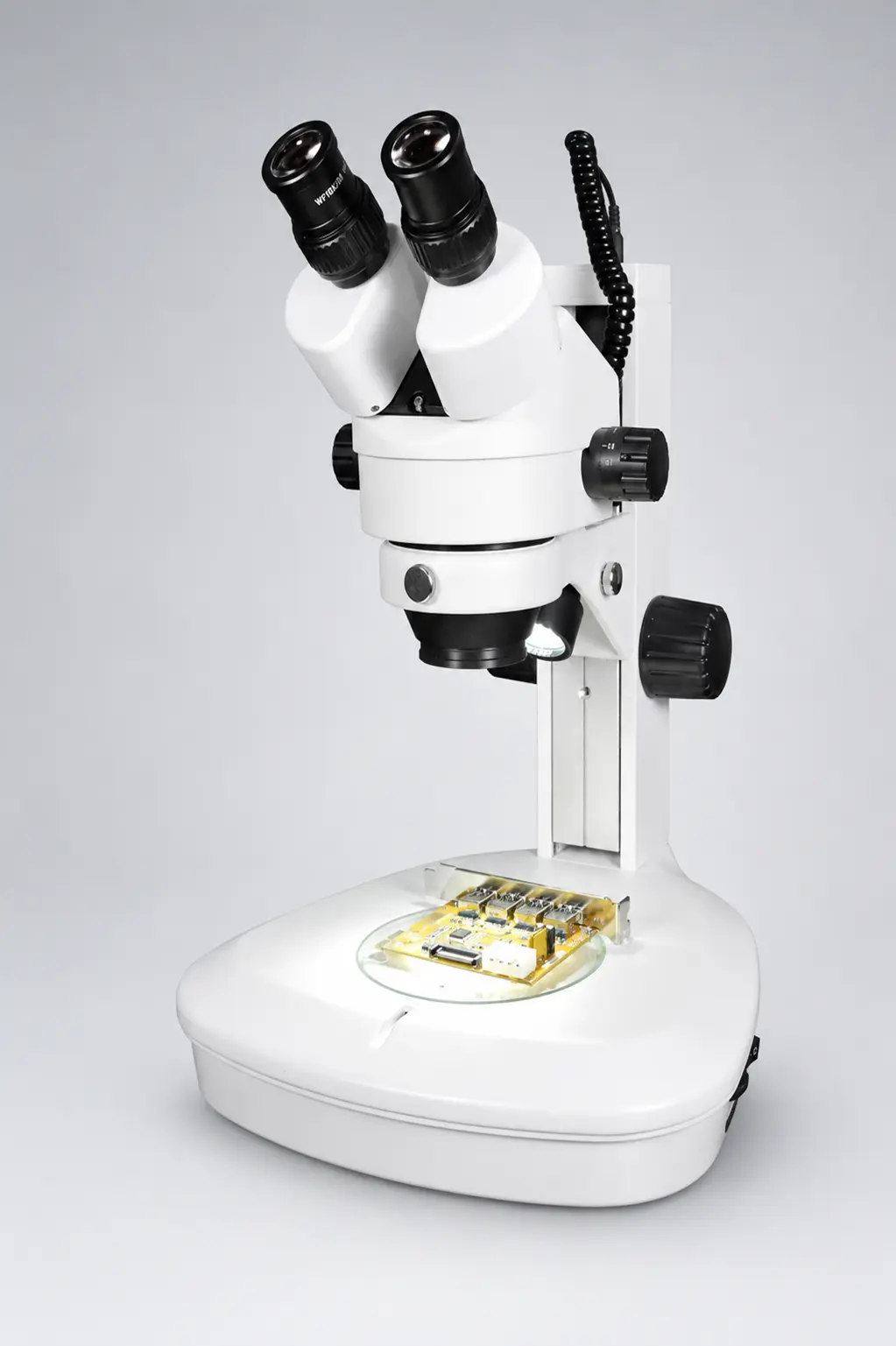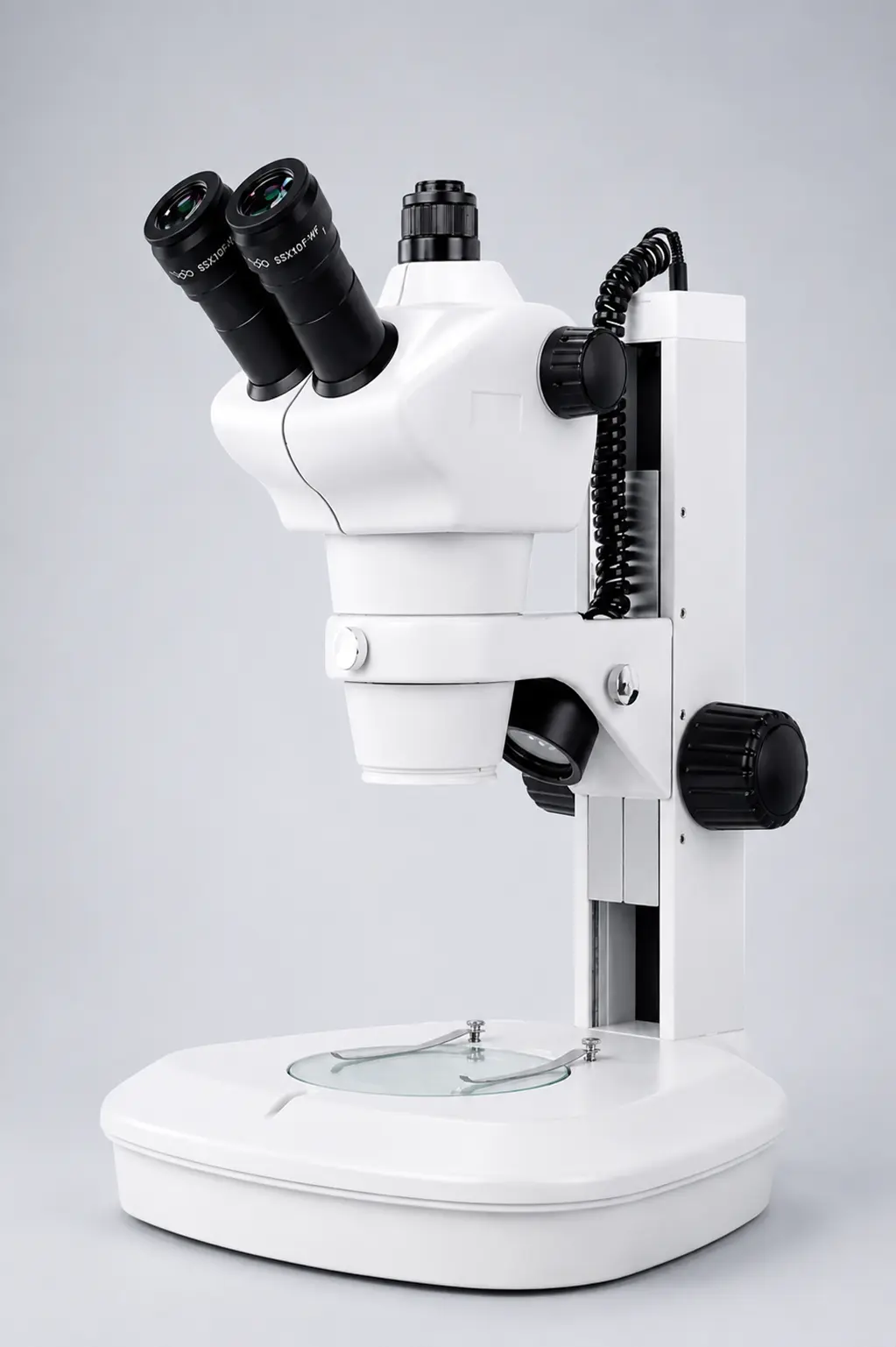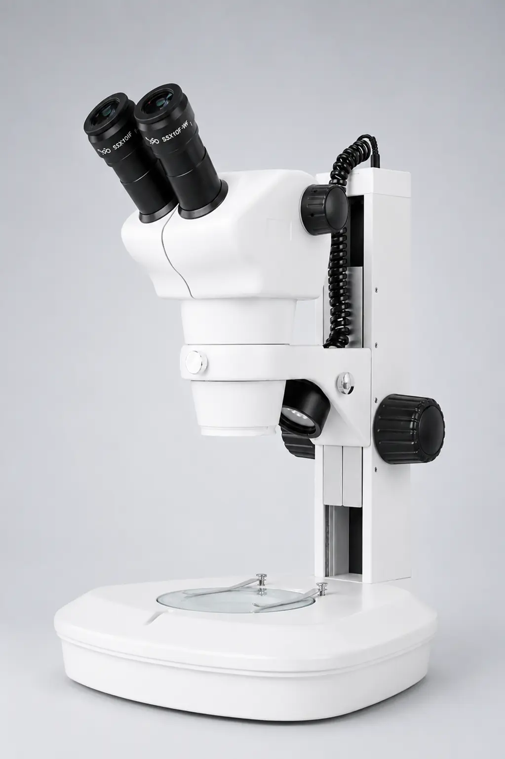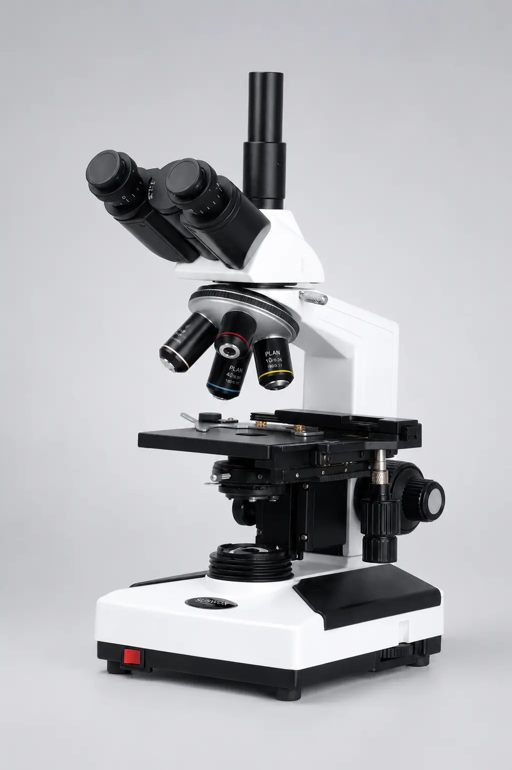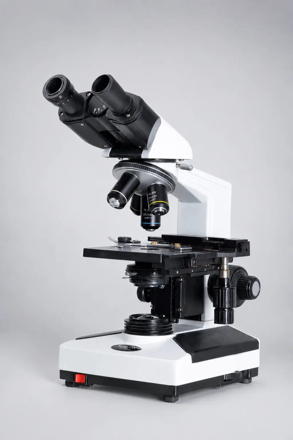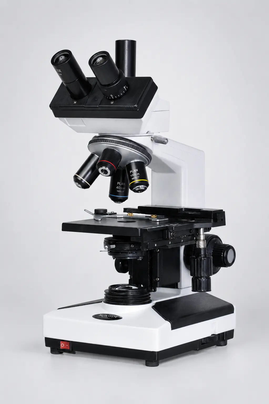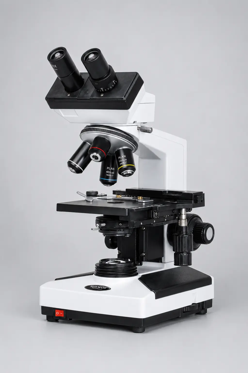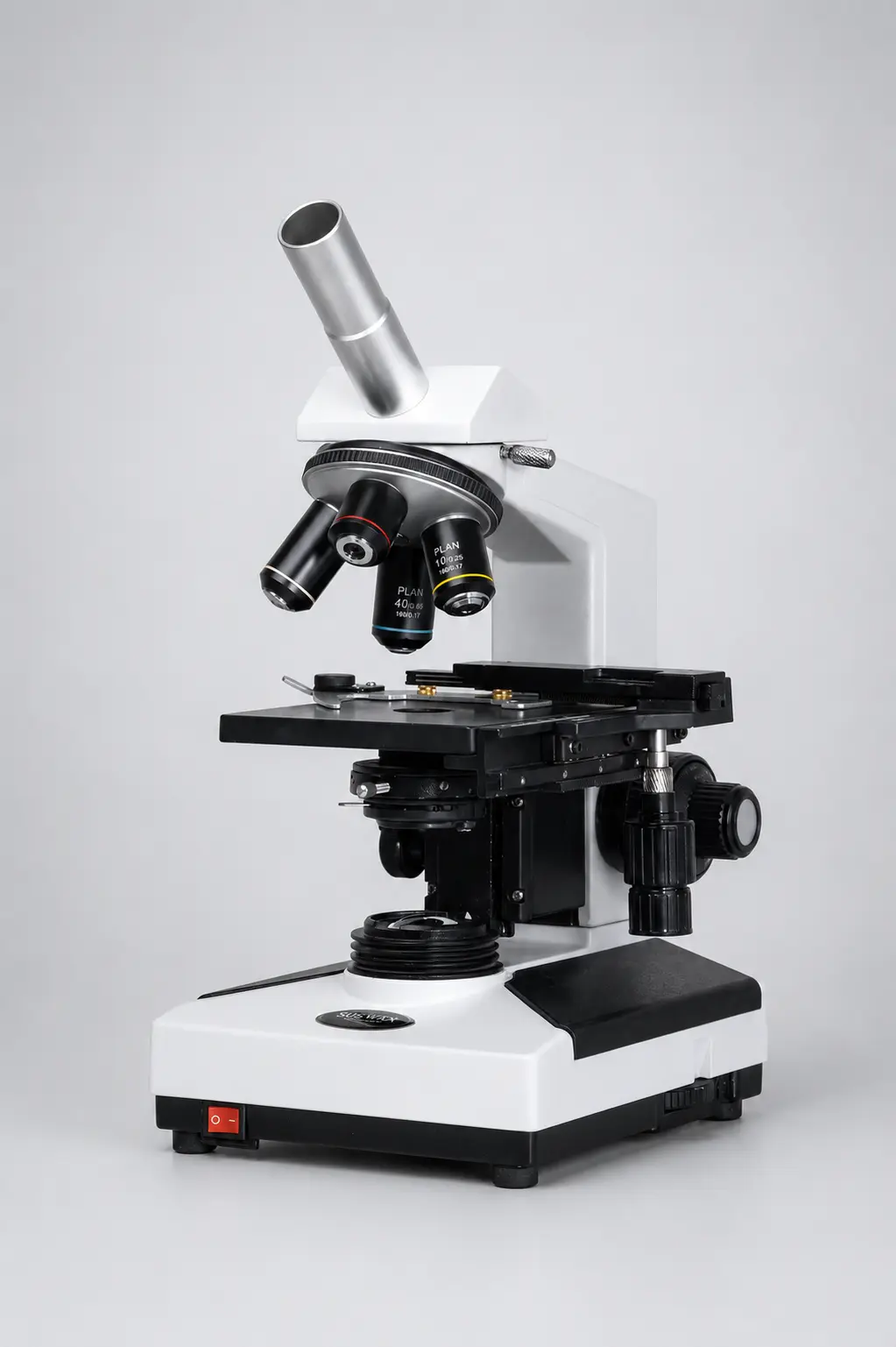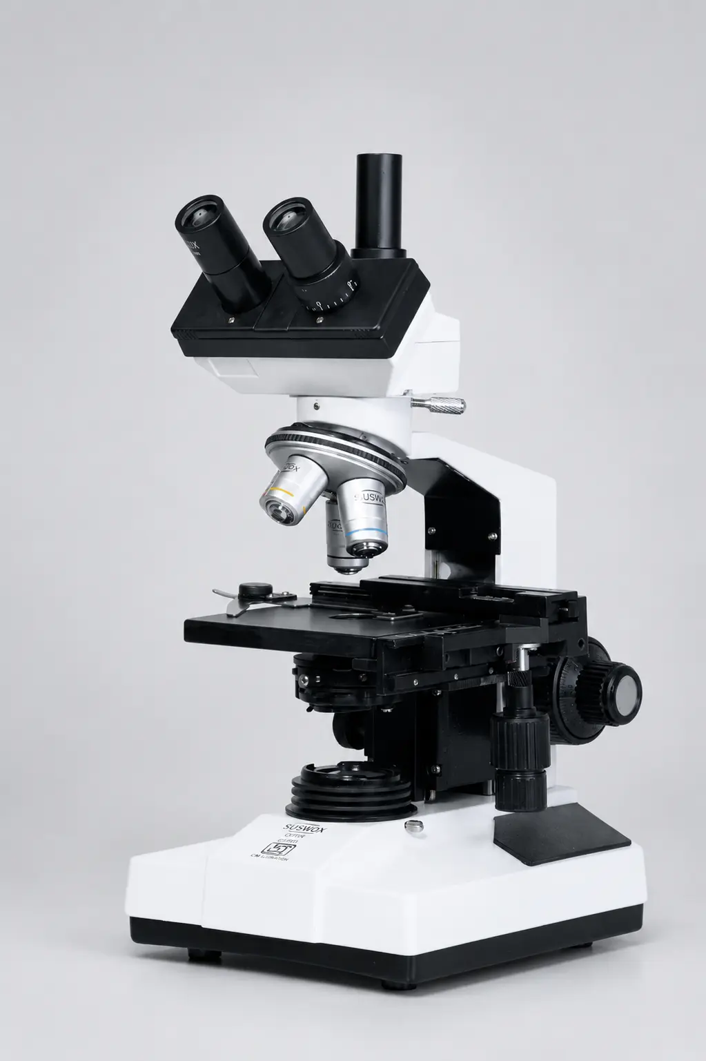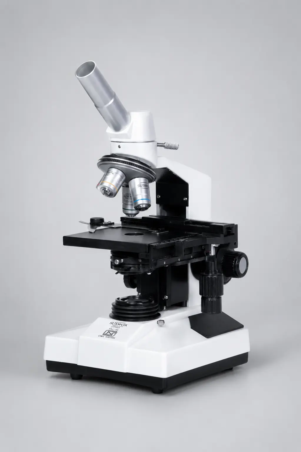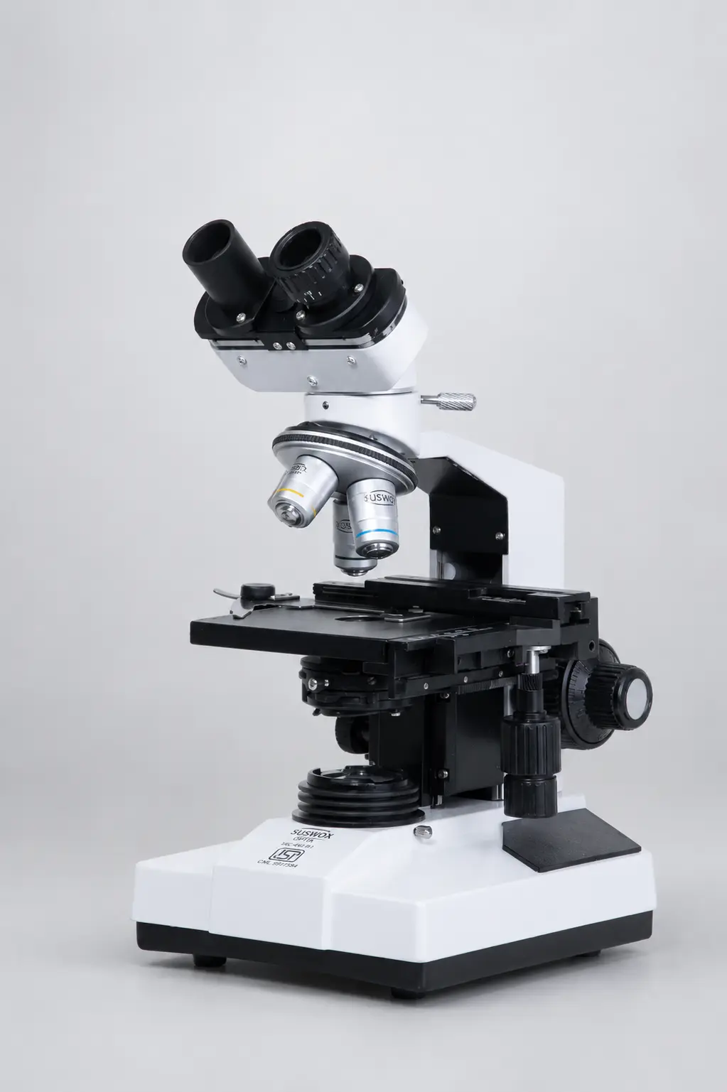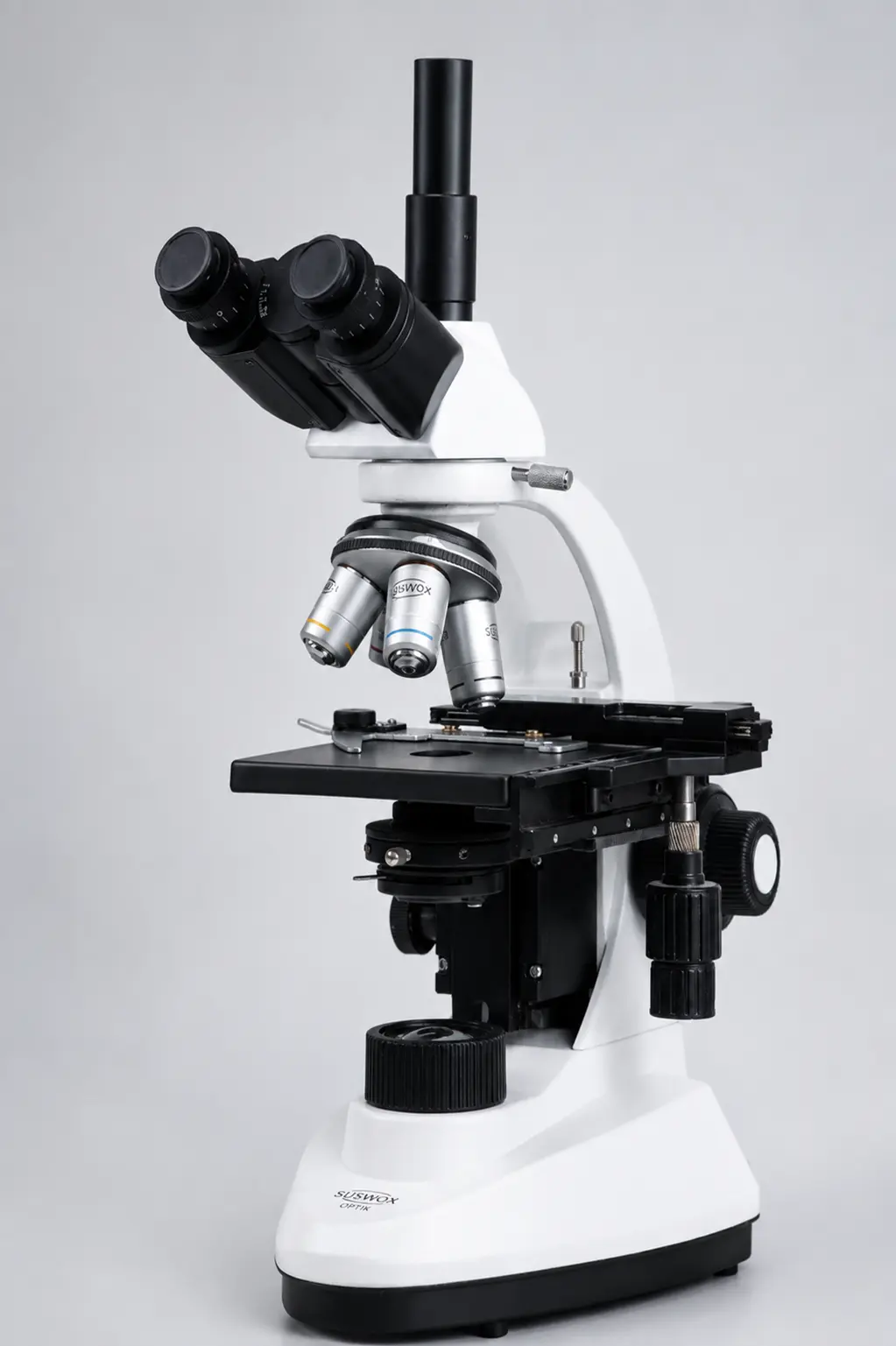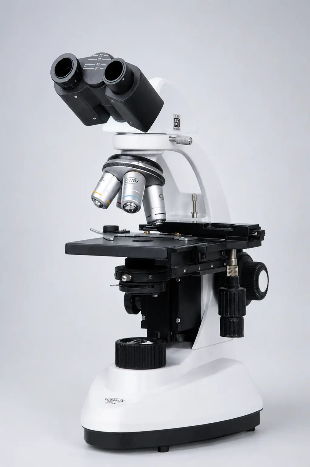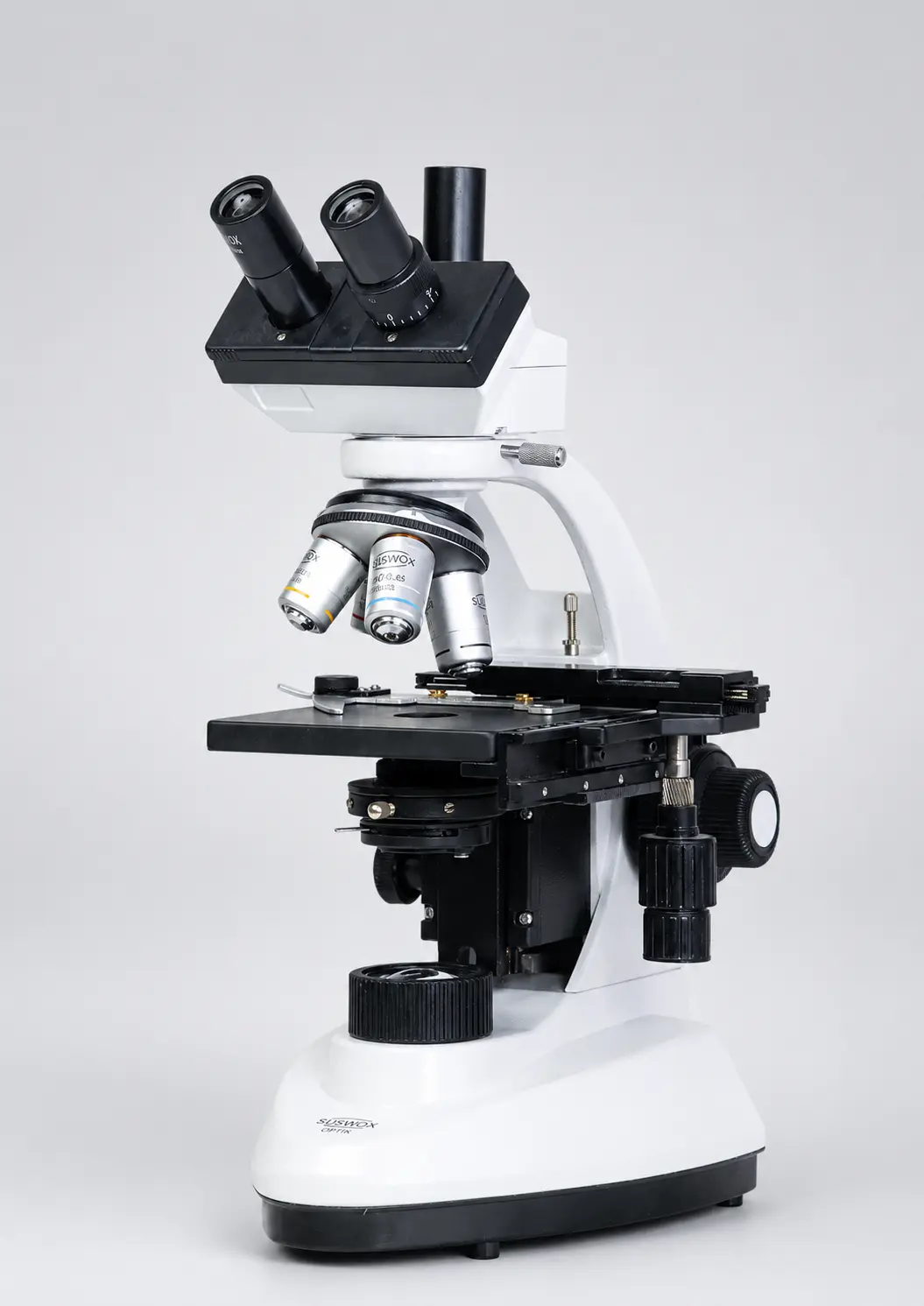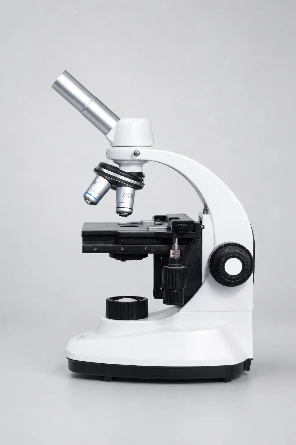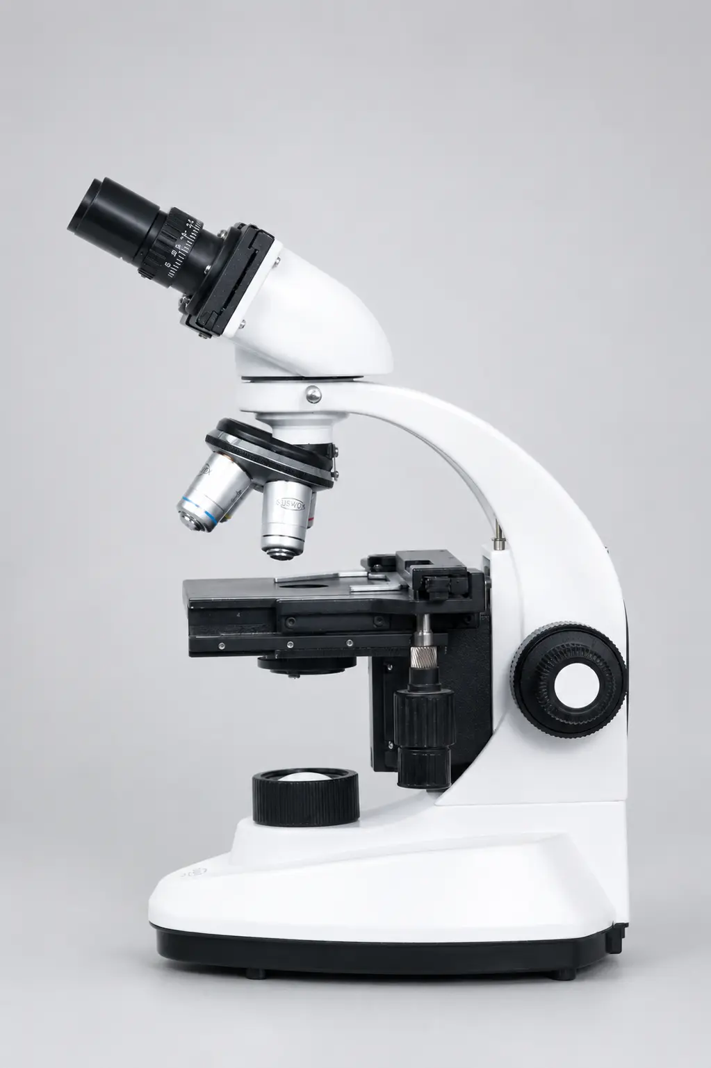
Put two microscopes side by side. Both marked 1000x. Both showing the same bacterial smear.
Through the first one, you can see individual rod-shaped cells with clean edges. Through the second, the same field looks like grey soup.
The magnification is identical. What separates them is resolving power — and it is the one specification most buyers never look at.
What is the resolving power of a microscope?
The resolving power of a microscope is its ability to show two very close points as two separate points instead of one merged blur. It is measured as the smallest distance between those two points at which they can still be told apart, and it depends on the wavelength of light used and the numerical aperture of the objective lens.
So a lower number is better. A microscope that resolves 0.2 µm is sharper than one that resolves 0.5 µm, even if both carry the same magnification figure on the box.
Why resolution beats magnification, every time
Magnification only makes the image larger. Resolution decides whether that larger image contains anything new.
Blow up a low-quality photo on your phone and it does not become clearer — it becomes a bigger version of the same blur. Microscopes behave exactly the same way. Push past what the optics can resolve and you get what microscopists call empty magnification: more size, zero extra information.
| Feature | Magnifying power | Resolving power |
| What it does | Makes the image bigger | Makes fine detail visible |
| Depends on | Objective x eyepiece | NA and wavelength of light |
| Typical figure | 400x, 1000x | 0.2 µm to 1.5 µm |
| Better value is | Higher | Lower (smaller distance) |
| Can you fix it later | Yes, change eyepiece | No, it is set by the optics |
| Importance | Secondary | Primary |
This is why an instrument advertising 2000x with a cheap objective is not a better microscope. It is a bigger blur.
The resolving power of microscope formula
There are two formulas in common use, and the difference between them confuses a lot of students.
| Formula | Expression | When it is used |
| Rayleigh criterion | d = 0.61 λ / NA | Standard for point-object resolution; most textbooks and lab work |
| Abbe limit | d = λ / (2 × NA) | Diffraction limit for periodic detail, gives a slightly smaller d |
| Abbe with condenser | d = λ / (NA objective + NA condenser) | Most realistic for bright-field work, since the condenser matters too |
| Resolving power | RP = 1 / d | The reciprocal — higher number means better |
Here, d is the resolution limit in the same unit as λ, λ is the wavelength of the light, and NA is the numerical aperture of the objective.
One point worth being careful about. Resolving power expressed as 1/d has units of “per micrometre” or “per nanometre” — not nanometres. You will see this written incorrectly in plenty of places online.
Breaking the formula into its two parts
Wavelength (λ)
Shorter wavelength, better resolution. That is the whole relationship.
| Light source | Wavelength | Resolution at NA 1.25 |
| Red | ~650 nm | ~0.32 µm |
| Green | ~550 nm | ~0.27 µm |
| Blue | ~450 nm | ~0.22 µm |
| Ultraviolet | ~350 nm | ~0.17 µm |
This is exactly why green filters are standard on bright-field microscopes. The eye is most sensitive to green, and the shorter wavelength improves resolution compared with plain warm-white light.
Numerical aperture (NA)
NA measures how wide a cone of light the objective can collect. The formula is:
NA = n × sin θ
where n is the refractive index of the medium between lens and specimen (air = 1.00, immersion oil = 1.515) and θ is the half-angle of the light cone entering the lens.
Wider cone, more diffracted light captured, finer detail resolved.
What your objectives actually resolve
Theory is fine. Here are the numbers for standard achromatic objectives at 550 nm green light, using the Rayleigh formula.
| Objective | Typical NA | Resolution (d) | Useful magnification range |
| 4x | 0.10 | ~3.4 µm | 50x – 100x |
| 10x | 0.25 | ~1.34 µm | 125x – 250x |
| 40x dry | 0.65 | ~0.52 µm | 325x – 650x |
| 100x oil | 1.25 | ~0.27 µm | 625x – 1250x |
That last column comes from a practical rule every lab should know: useful magnification sits between 500 and 1000 times the NA. Below 500 × NA you are throwing away detail the lens could show you. Above 1000 × NA you are in empty magnification.
So a 40x/0.65 objective with a 10x eyepiece gives 400x — comfortably inside its useful range. The same objective with a 25x eyepiece gives 1000x, which is well past its limit and looks worse, not better.
Why oil immersion works
Between a coverslip and a dry objective sits a layer of air. Light travelling from glass (n = 1.52) into air (n = 1.00) bends sharply away, and the widest-angle rays — the ones carrying the finest detail — never reach the lens at all.
That is a hard ceiling. A dry objective cannot exceed NA 1.0, and in practice tops out around 0.95.
Fill that gap with immersion oil at n = 1.515, almost identical to glass, and the light travels straight through without bending away. NA rises to 1.25 or 1.30, and resolution improves by roughly half.
Which is also why using an oil objective without oil gives a poor image. The design assumes the oil is there.
The part most guides skip: your condenser
Here is where theory and the bench part company.
The Abbe formula includes the condenser NA for a reason — the condenser illuminates the specimen, and if it delivers a narrow cone of light, the objective has nothing wide to collect. Closing the condenser iris to increase contrast is the single most common way labs quietly throw away resolution.
Working through it: with a 1.25 NA objective and a 0.90 NA condenser at 550 nm, real resolution is about 0.26 µm. Stop the condenser iris down to an effective 0.30 and it collapses to around 0.35 µm. Same microscope, same lens, one-third of the detail gone.
Set the condenser properly using Köhler illumination and open the iris to about 70–80% of the objective NA. It costs nothing and it is the biggest free improvement available on most instruments.
Worked examples
Example 1 — oil immersion, green light. NA = 1.25, λ = 550 nm.
d = 0.61 × 550 / 1.25 = 268 nm ≈ 0.27 µm
Example 2 — high-power dry lens, blue light. NA = 0.90, λ = 450 nm.
d = 0.61 × 450 / 0.90 = 305 nm ≈ 0.31 µm
Example 3 — resolving power as a reciprocal. Taking d = 268 nm from Example 1:
RP = 1 / 268 nm ≈ 0.0037 per nm, or about 3.7 lines per micrometre.
Notice that Example 2 uses a dry lens with shorter wavelength and still cannot beat oil immersion with green light. NA moves the number more than wavelength does.
Resolution limits across microscope types
| Microscope type | Approximate resolution limit |
| Human eye | ~100 µm |
| Bright-field light microscope | ~200 nm (0.2 µm) |
| Confocal | ~150–180 nm |
| Super-resolution (STED, PALM) | ~20–50 nm |
| SEM | ~1–10 nm |
| TEM | ~0.1 nm |
The 200 nm figure for optical microscopes is a physical limit, not a manufacturing one. No amount of spending improves it while you are using visible light through conventional optics.
Practical ways to improve resolution
- Open the condenser iris to roughly 70–80% of the objective NA, and set up Köhler illumination properly
- Use oil correctly with 100x objectives — fresh oil, no bubbles, cleaned off after every session
- Switch to a green filter for routine bright-field work
- Use 0.17 mm coverslips, because high-NA objectives are computed for that exact thickness
- Cut thinner sections — thick specimens scatter light and destroy contrast before optics ever become the limit
- Clean the front element, since dried oil costs you more detail than any specification difference between brands
- Choose plan achromatic objectives if you photograph slides, so the field stays sharp to the edges
Common misconceptions
“Higher magnification means higher resolving power.” No. Resolution comes from NA and wavelength. A 2000x instrument with a 0.65 NA lens resolves exactly what a 400x instrument with the same lens resolves.
“Only the objective matters.” The condenser, the light source, the coverslip thickness and the mounting medium all sit in the optical path.
“The same formula applies to electron microscopes.” Electron microscopes use electron wavelength, which is thousands of times shorter, and follow different aberration limits.
FAQs
What is the resolving power of a microscope in simple words?
It is the smallest gap between two points at which a microscope can still show them as two separate points. Smaller gap, better instrument.
What is the resolving power of microscope formula?
The Rayleigh form is d = 0.61 λ / NA, giving the resolution limit. Resolving power itself is the reciprocal, RP = 1 / d. The Abbe form, d = λ / 2NA, is also widely used.
What is the maximum resolution of a light microscope?
About 200 nm, or 0.2 µm, using visible light and an oil immersion objective. This is a diffraction limit set by physics.
Does a higher eyepiece magnification improve resolution?
No. It only enlarges what the objective has already resolved. Past 1000 × NA, the extra magnification is empty.
How does numerical aperture affect resolution?
Resolution improves in direct proportion to NA. Doubling NA halves d, which is why oil immersion at 1.25 outperforms a dry lens at 0.65 so clearly.
Why does my microscope look blurry even at 1000x?
Usually a closed condenser iris, missing or dirty immersion oil, a wrong coverslip thickness, or a thick specimen — long before the optics are actually at fault.
Choosing optics that deliver what the formula promises
Every number in this article assumes the objective actually performs to its printed NA. That is where manufacturing quality stops being theoretical.
When comparing instruments, ask for the objective type and NA value of every lens, whether the condenser is adjustable with an iris, and what illumination is fitted. A supplier confident in its optics will give you those figures without hesitation. One that only quotes total magnification is telling you the least useful number available.
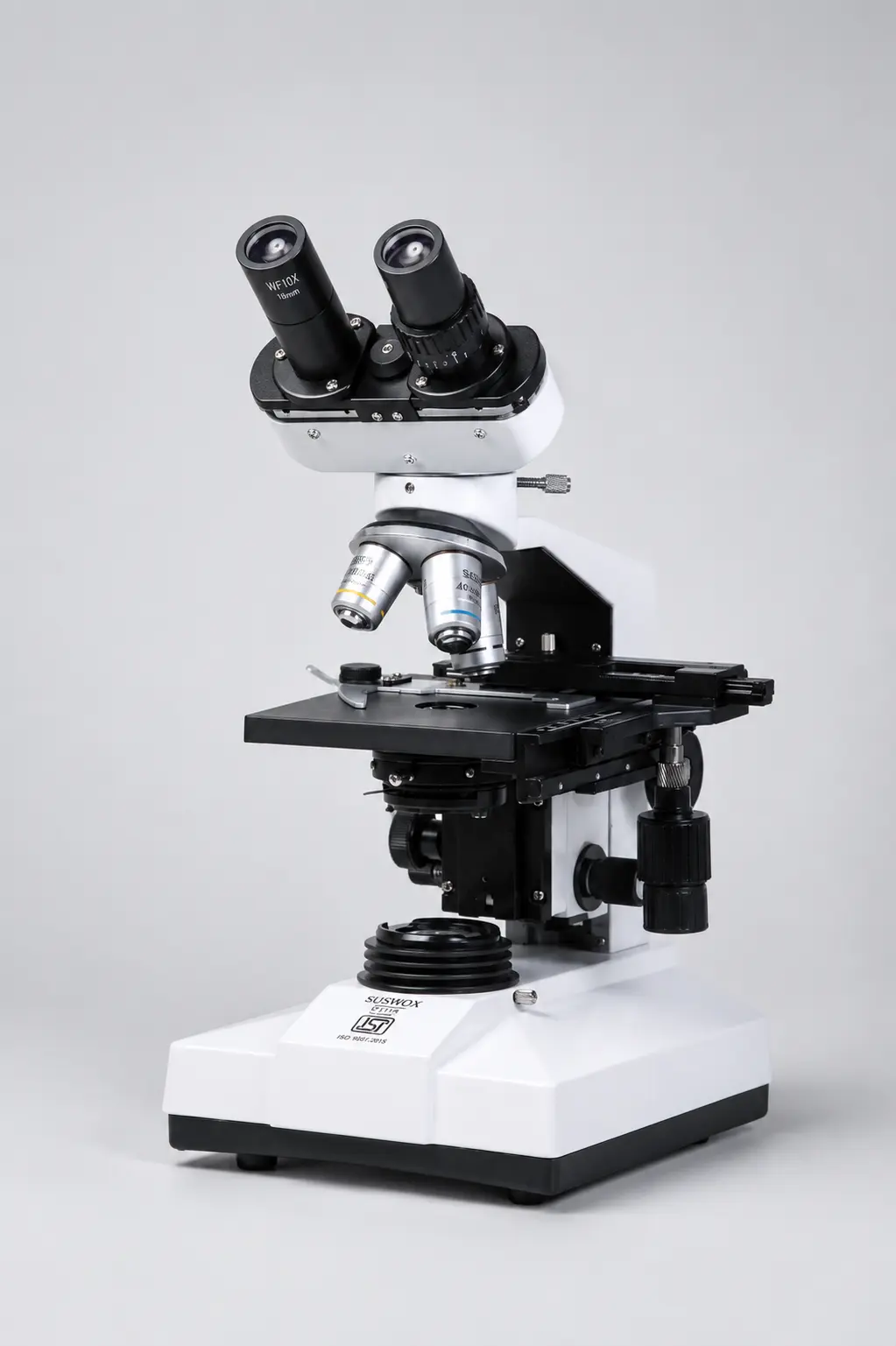
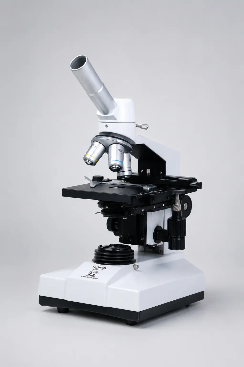
SUSWOX, the microscopy brand of Sudheer Scientific Works, has been manufacturing ISI-marked optical instruments in Ambala since 1973 — compound, stereo zoom, inverted, digital, travelling and projection models, all assembled and quality-checked in-house. Instruments are in daily use at AIIMS Nagpur, Bathinda and Bilaspur, BHU, MDU Rohtak and the Haffkine Institute, with spares and servicing supported long after delivery.
If you are comparing options as a microscope manufacturer in India, start with the NA values and the condenser specification. Those two numbers tell you what you will actually see through the eyepiece.