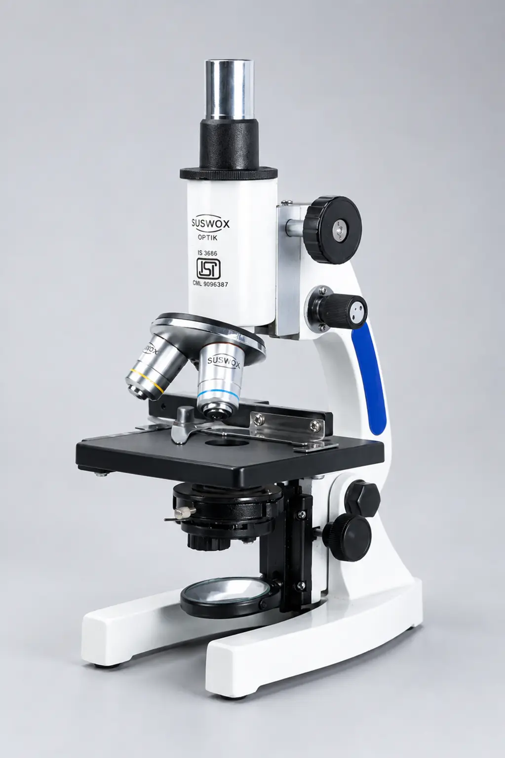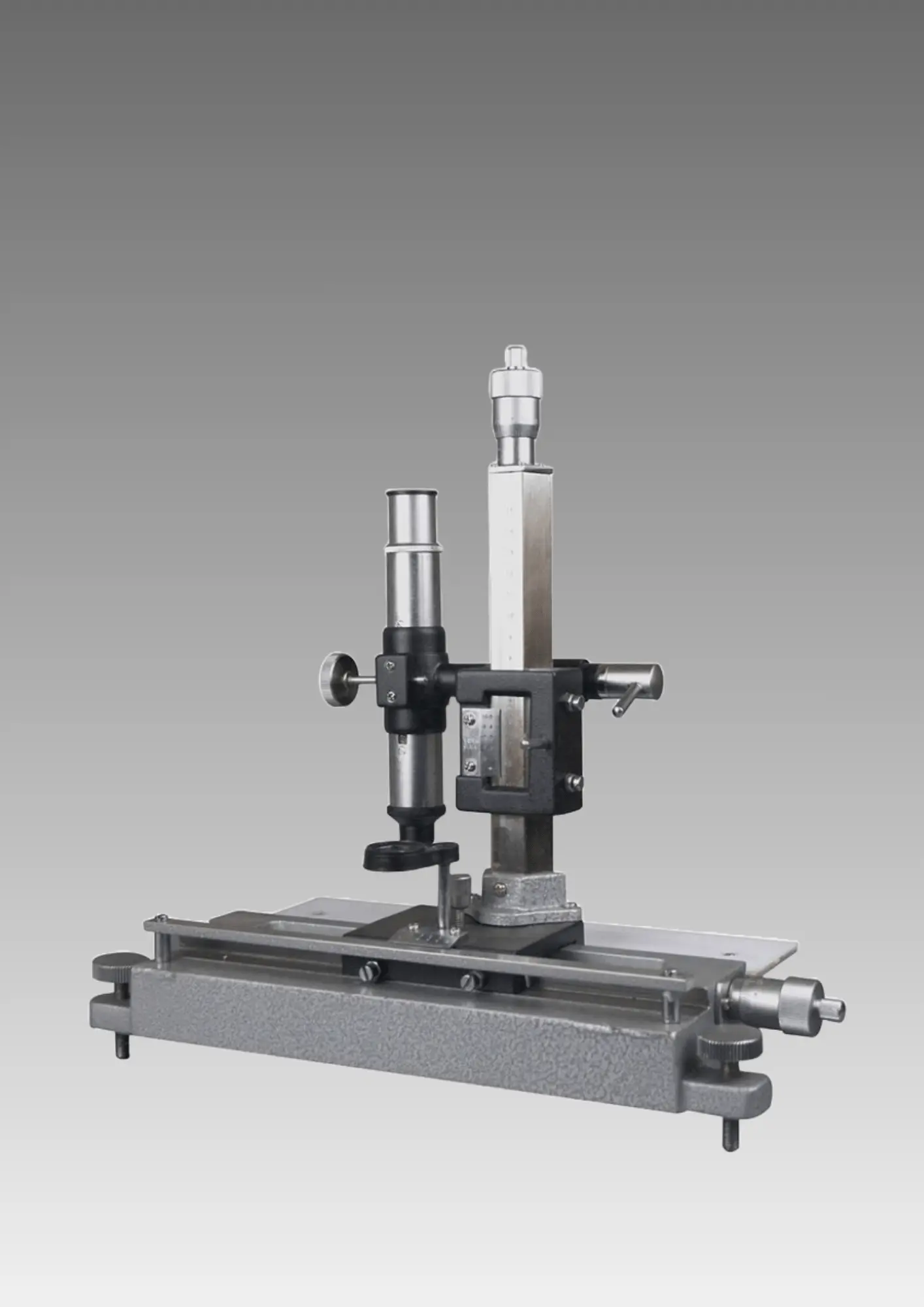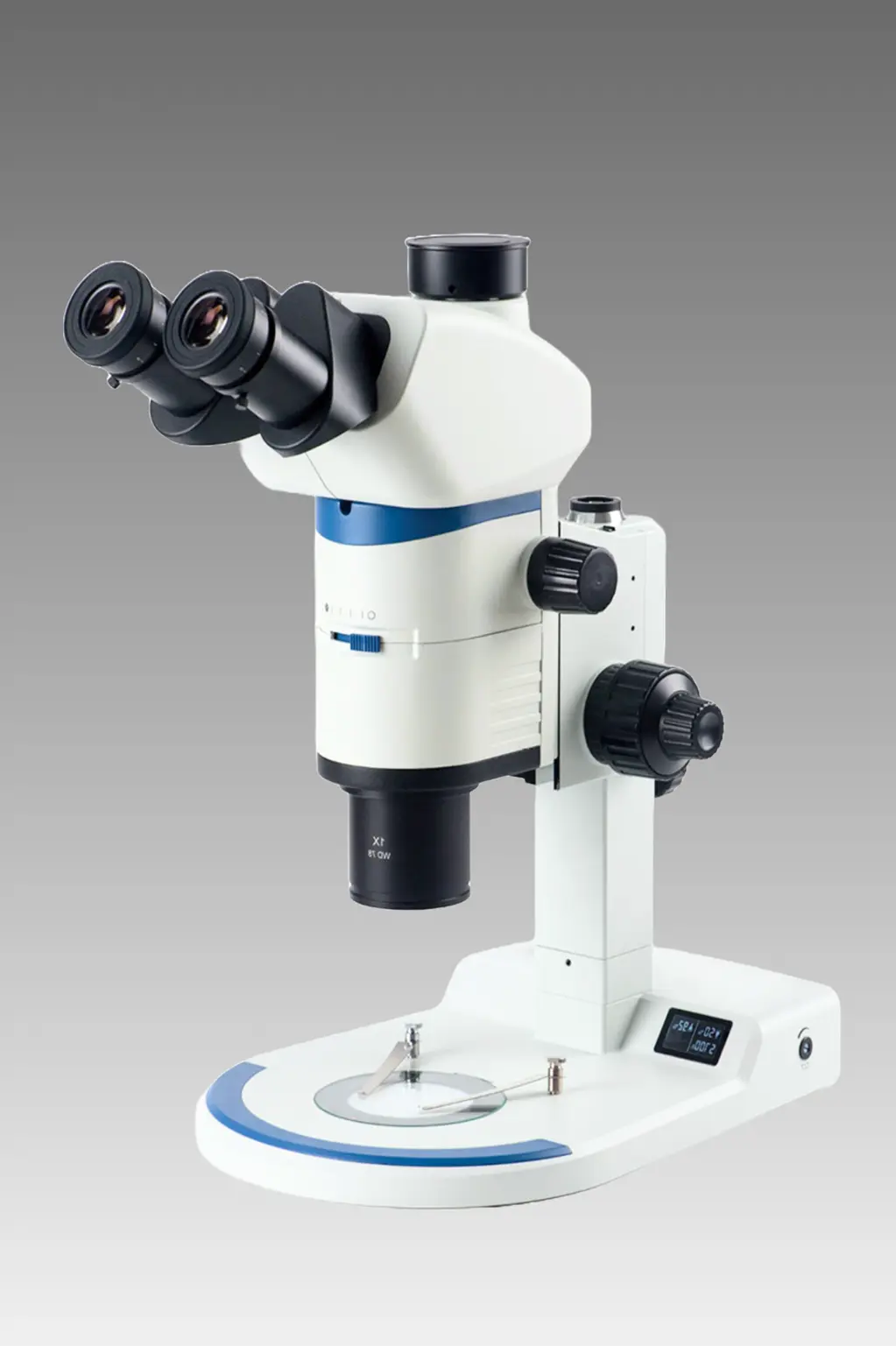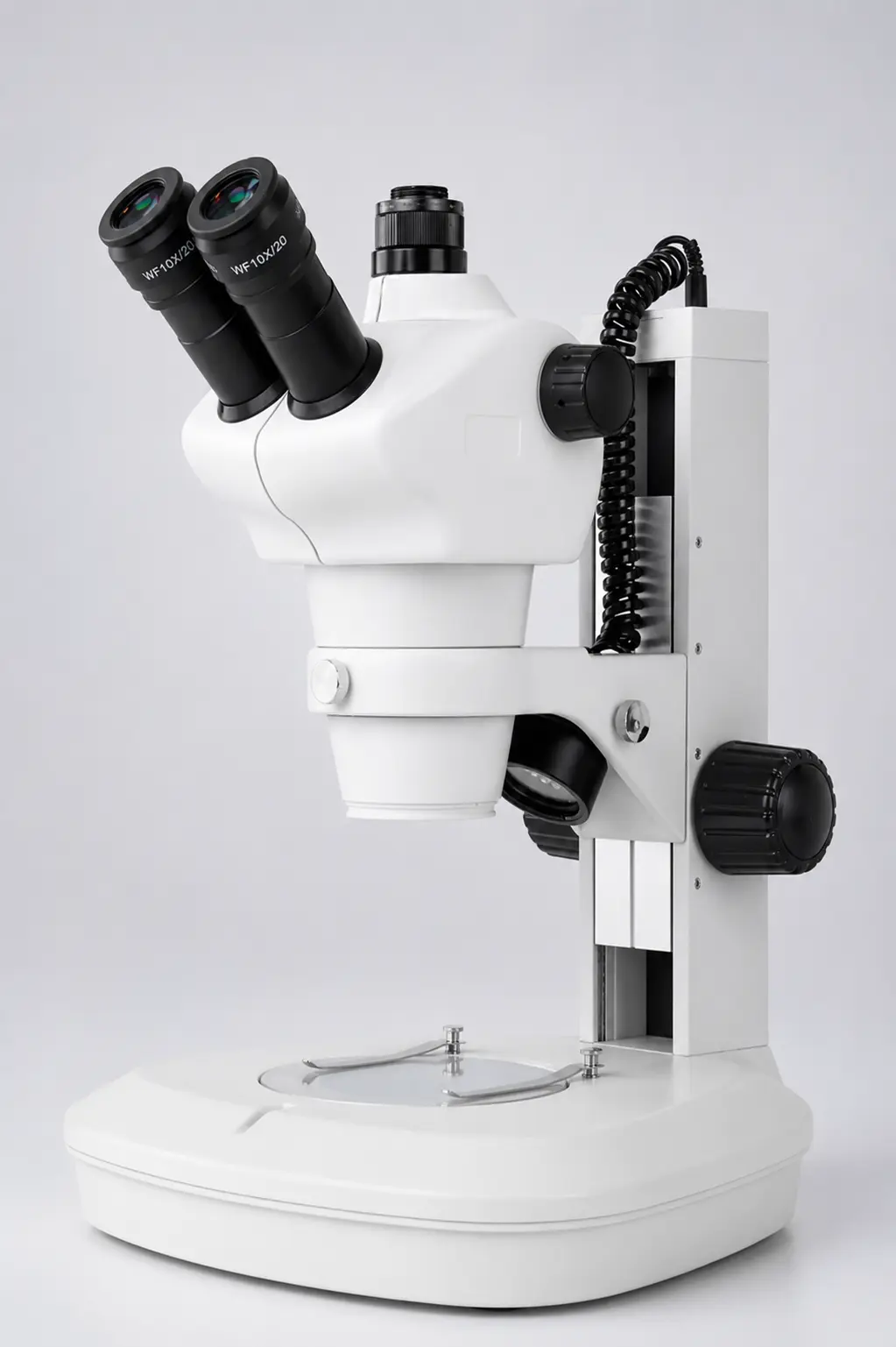
A physics teacher in a Haryana government college once ordered nine microscopes for her lab. Good budget. Proper approval. Everything cleared.
And then the crates arrived, and every single one of them was a compound biological microscope — while half her practicals that semester needed a travelling microscope to measure refractive index.
That mix-up is more common than you would think.
The word “microscope” covers dozens of very different instruments, and picking the wrong category is an expensive mistake. So this guide walks through the 12 main types of microscopes, what each one actually does, where it is used, and roughly what it costs in India in 2026.
What are the different types of microscopes?
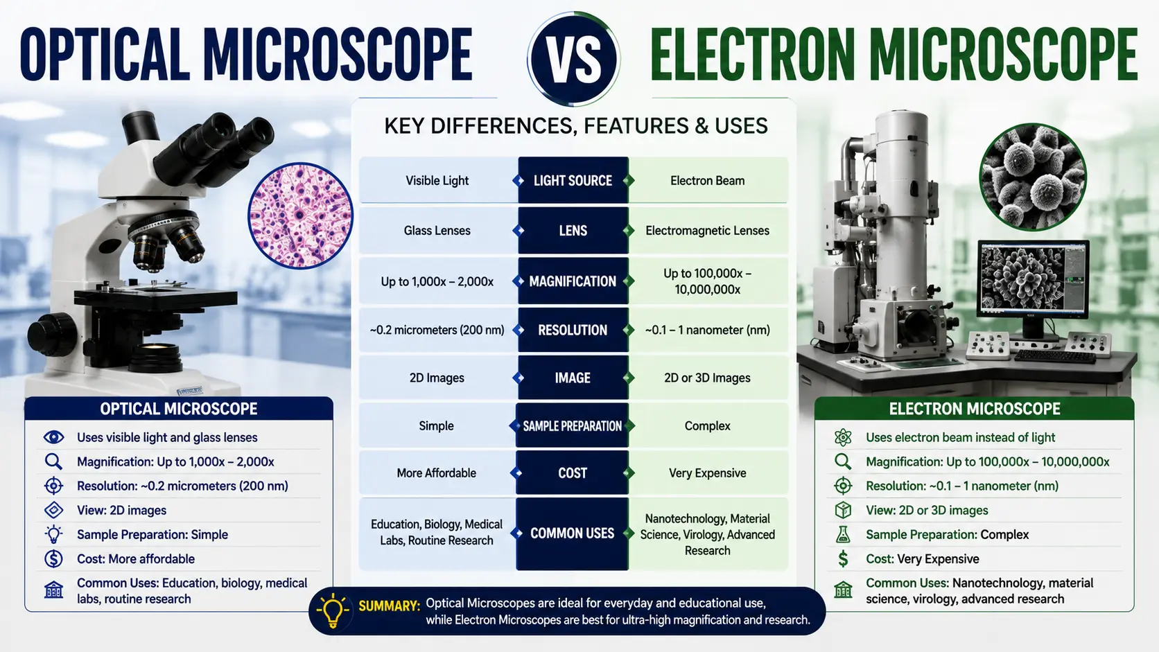
There are two broad types of microscopes: optical (light) microscopes and electron microscopes. Optical microscopes use visible light and glass lenses, and include simple, compound, stereo, inverted, digital, travelling, polarizing and fluorescence models. Electron microscopes use electron beams instead of light, and include SEM and TEM.
Everything else is a variation of those two families.
Quick Comparison of All 12 Types of Microscopes
Bookmark this table. It answers most questions before you even scroll down.
| # | Type of Microscope | Magnification | Image Type | Mainly Used In | Approx. Price in India (2026) |
| 1 | Simple Microscope | 5x – 100x | 2D, erect | Schools, jewellery, field study | ₹200 – ₹3,000 |
| 2 | Compound Microscope | 40x – 1000x | 2D, inverted | Pathology, biology, research | ₹8,000 – ₹1,50,000 |
| 3 | Student Microscope | 40x – 675x | 2D, inverted | Schools, colleges, coaching labs | ₹3,000 – ₹15,000 |
| 4 | Stereo Zoom Microscope | 6.5x – 90x | 3D, erect | Electronics, PCB, dissection | ₹12,000 – ₹80,000 |
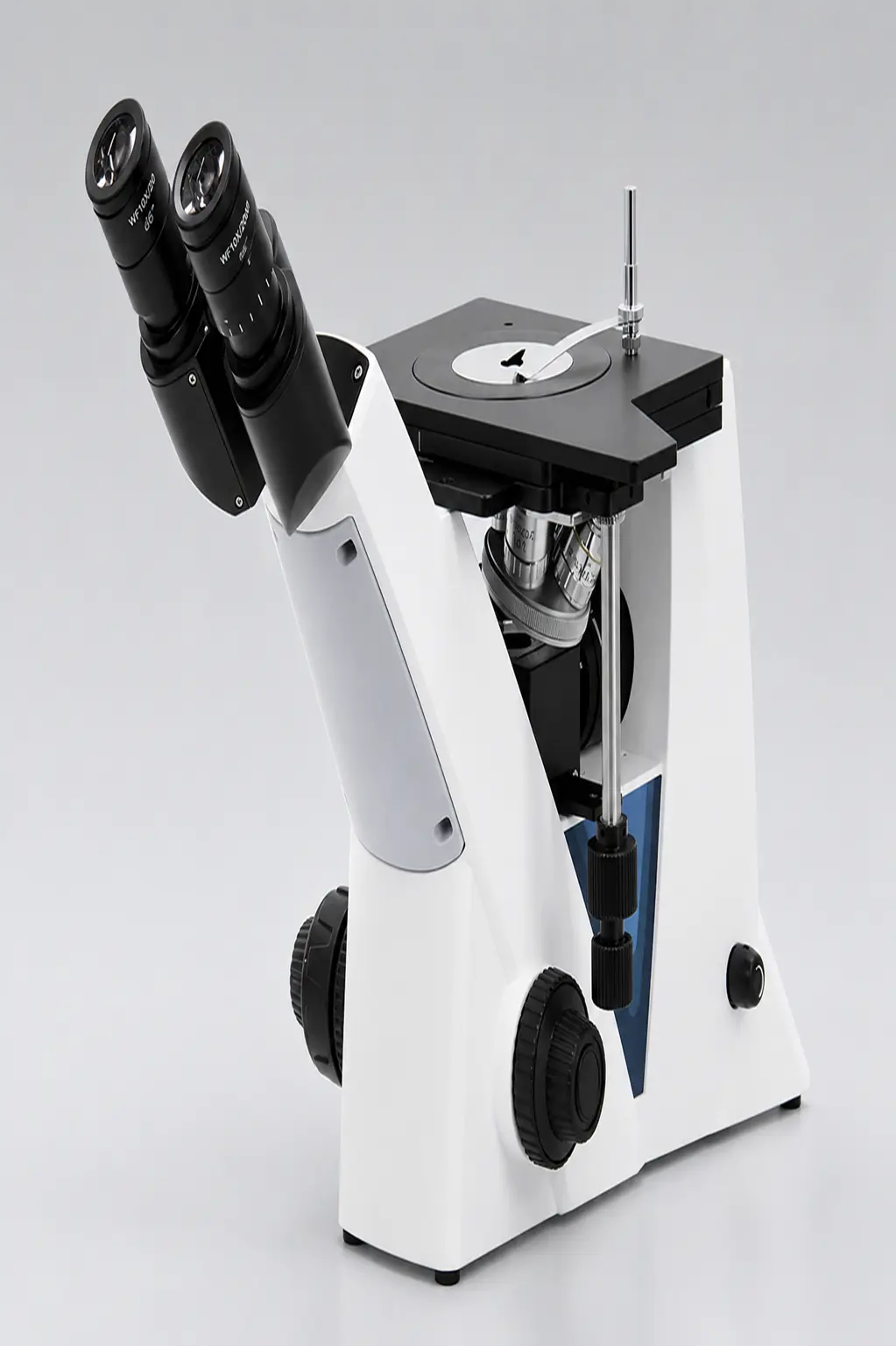
| 5 | Inverted Microscope | 40x – 400x | 2D, inverted | Tissue culture, IVF, cell work | ₹60,000 – ₹4,00,000 |
| 6 | Digital Microscope | 20x – 1000x | On-screen | QC, training, documentation | ₹5,000 – ₹1,20,000 |
| 7 | Travelling Microscope | 10x – 20x (with vernier) | 2D, erect | Physics labs, metrology | ₹6,000 – ₹25,000 |
| 8 | Projection Microscope | 100x – 600x | Projected image | Classrooms, demonstrations | ₹15,000 – ₹60,000 |
| 9 | Operating Microscope | 4x – 40x | 3D, erect | ENT, dental, ophthalmic surgery | ₹1,50,000 – ₹25,00,000 |
| 10 | Polarizing Microscope | 40x – 600x | 2D with colour data | Geology, pharma, minerals | ₹45,000 – ₹3,50,000 |
| 11 | Fluorescence Microscope | 100x – 1000x | Glowing on dark field | Immunology, TB testing, research | ₹1,50,000 – ₹12,00,000 |
| 12 | Electron Microscope (SEM/TEM) | 1,000x – 10,00,000x | Greyscale digital | Nanotech, materials, virology | ₹40,00,000+ |
Prices are indicative ranges for Indian buyers in 2026 and change with optics grade, illumination and accessories. For an exact quote on any category, request a quote from Suswox.
1. Simple Microscope
What is a simple microscope?
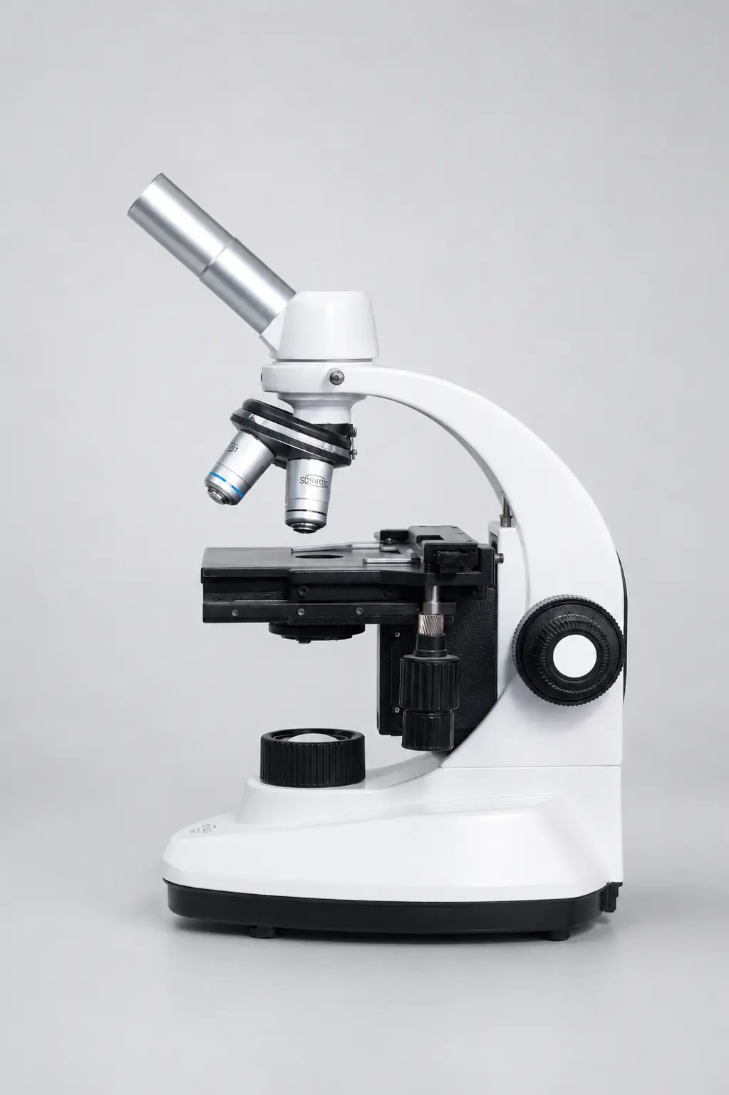
A simple microscope is a magnifying device that uses a single convex lens to produce an enlarged, erect and virtual image of an object. It magnifies between 5x and 100x, has no objective-eyepiece combination, and is the oldest and most basic of all types of microscopes.
Think of a hand lens. That is a simple microscope. So is a jeweller’s loupe.
Antonie van Leeuwenhoek built single-lens instruments in the 1670s that reached roughly 270x — far better than anything else in his era — and used them to become the first person to see bacteria. One lens. That’s it.
Where it is used today:
- Botany field work — checking leaf surfaces and stomata on the spot
- Jewellery and gemstone inspection
- Dermatology clinics for skin surface checks
- Watch repair and small mechanical assembly
- Primary school science introduction
Best for: basic magnification where portability matters more than power.
Limitation: no resolving power for cells. You will see a hair clearly. You will never see a bacterium.
[IMAGE 1 — file: simple-microscope-magnifying-lens.webp | alt: “Simple microscope with single convex lens used for basic magnification”]
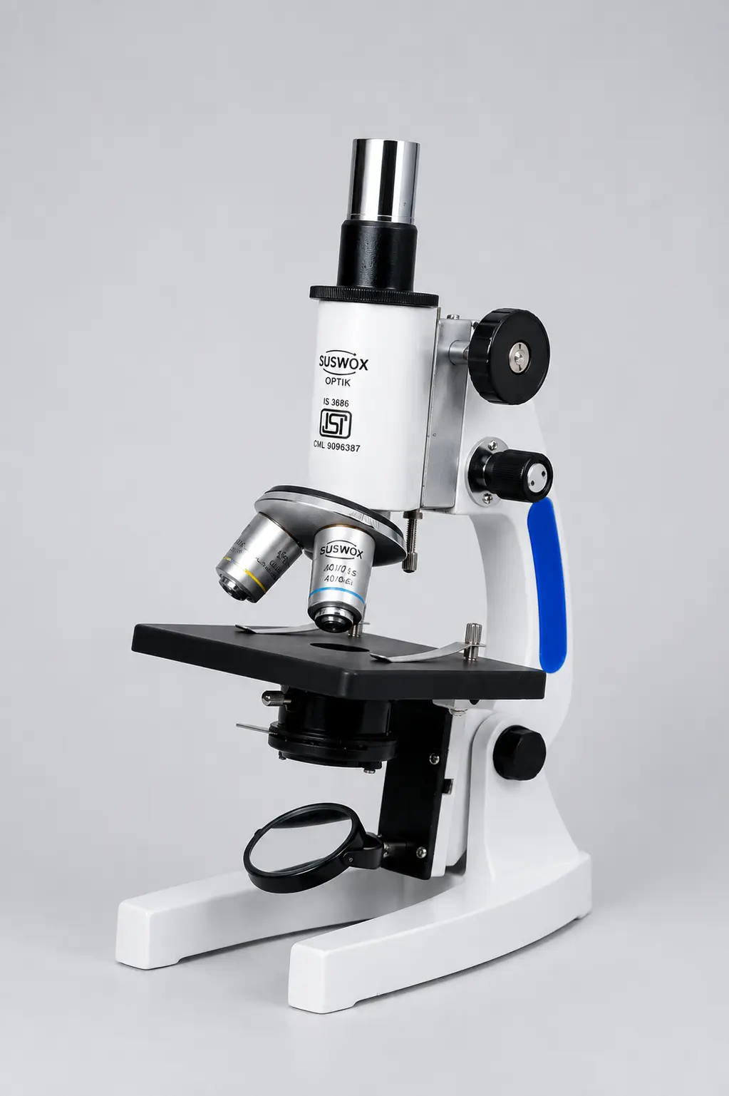
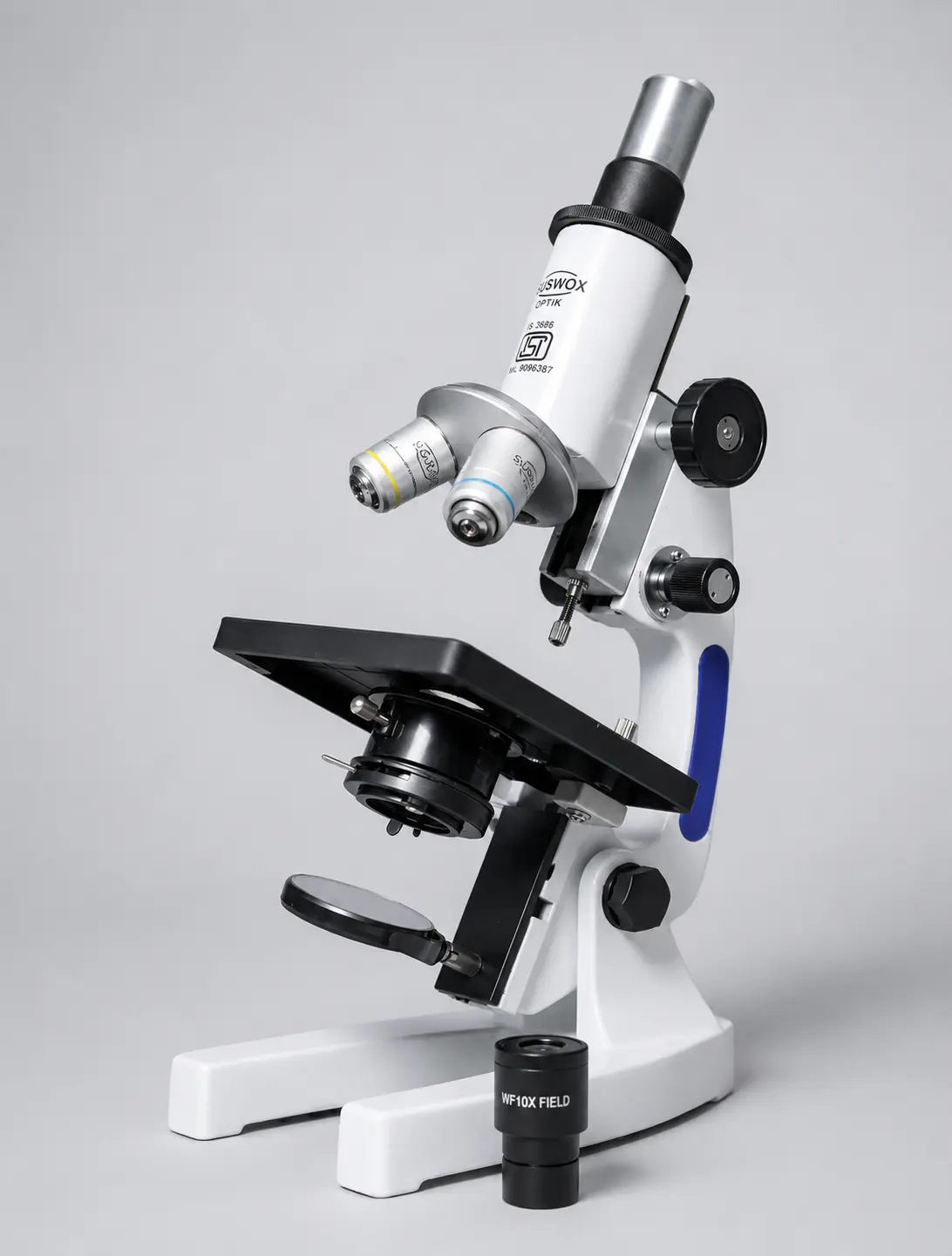
2. Compound Microscope
What is a compound microscope?
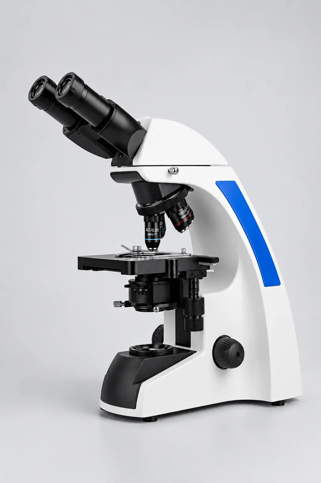
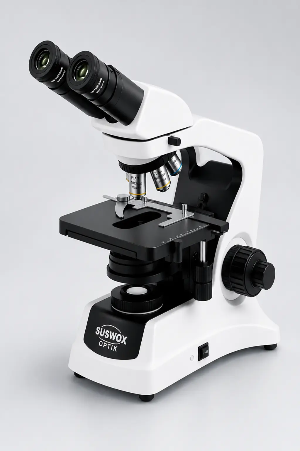
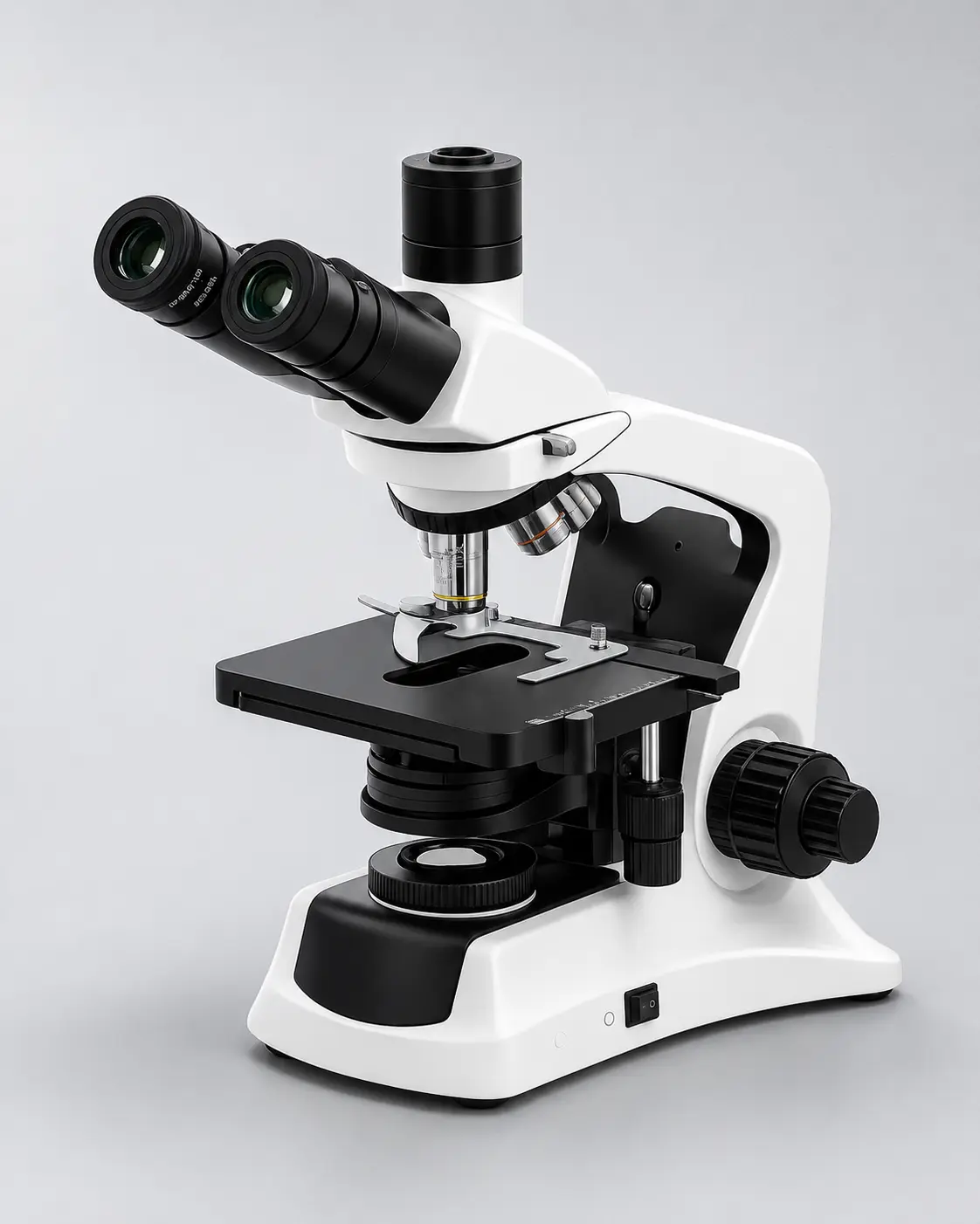
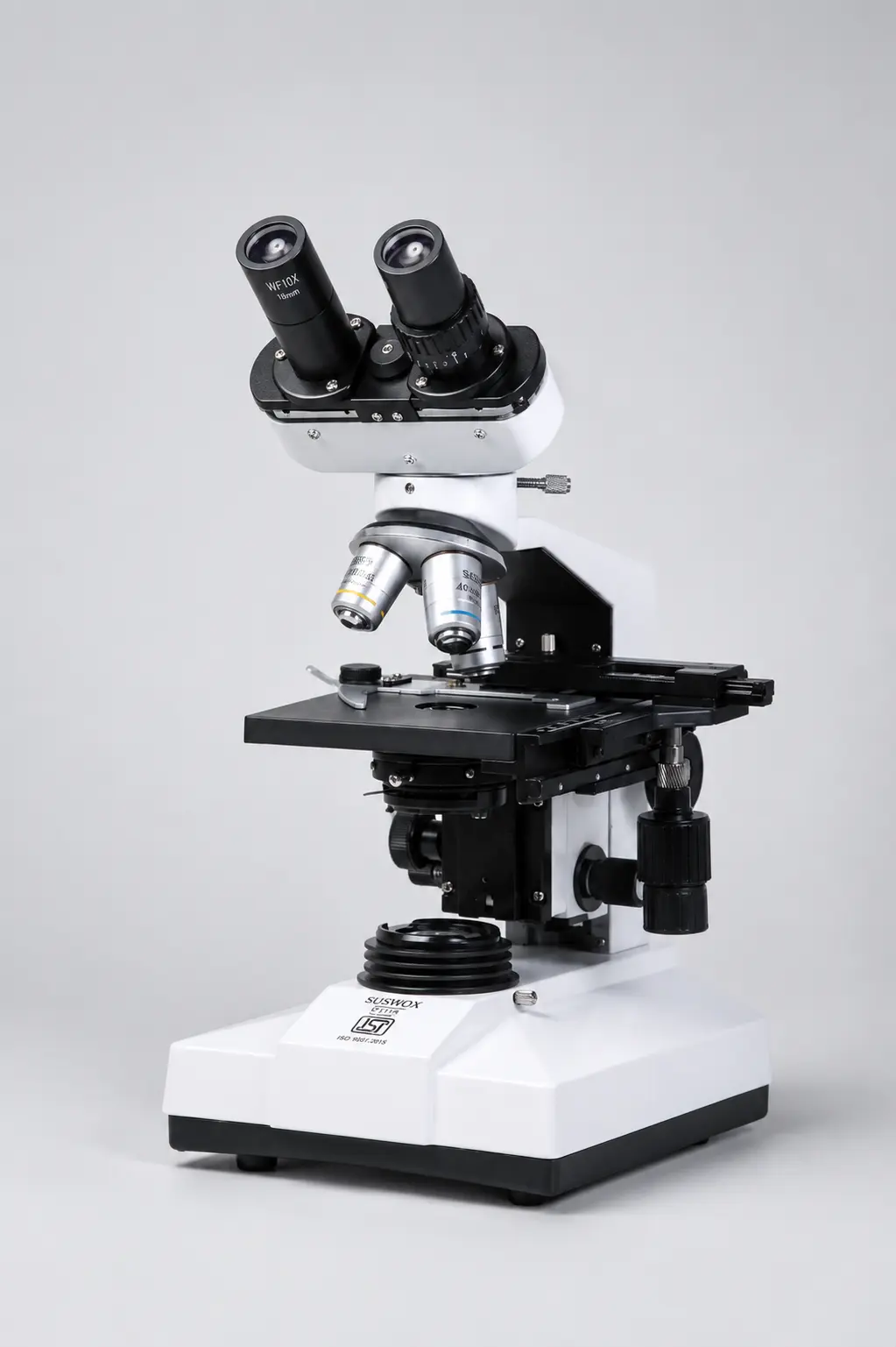
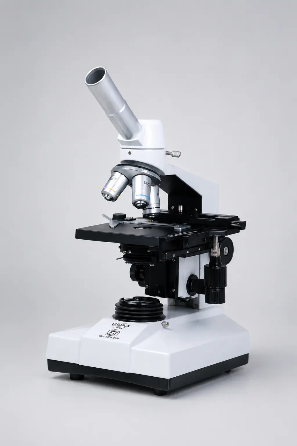
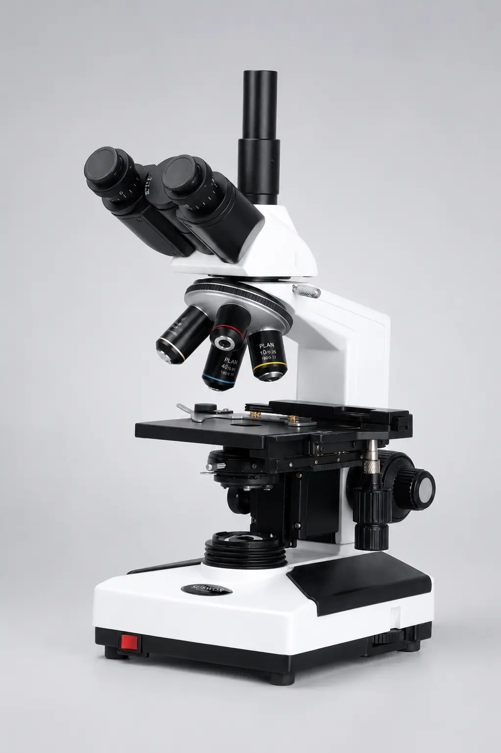
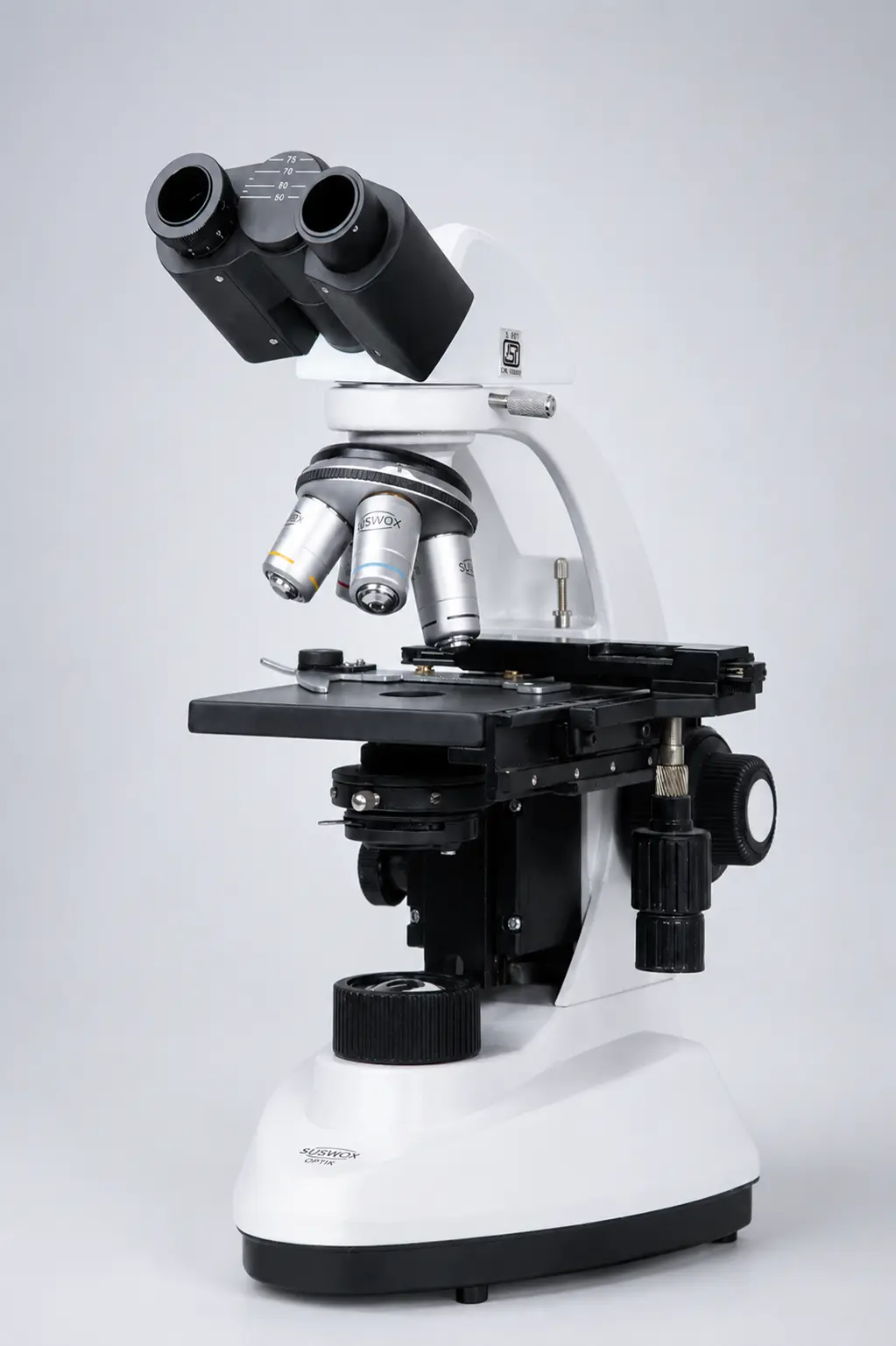
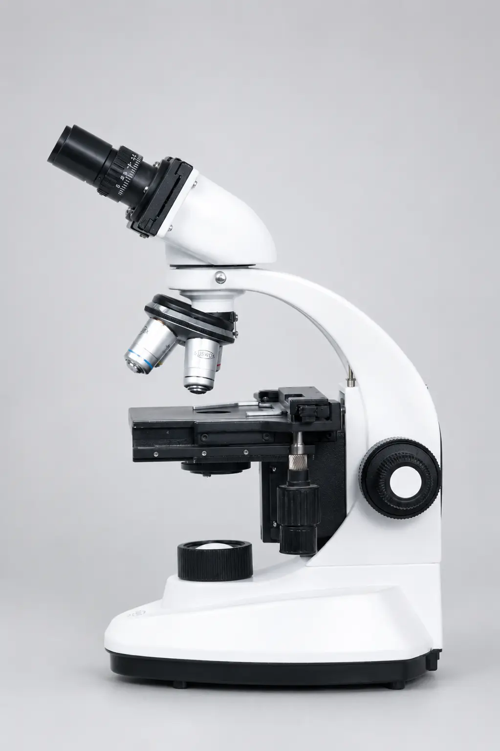
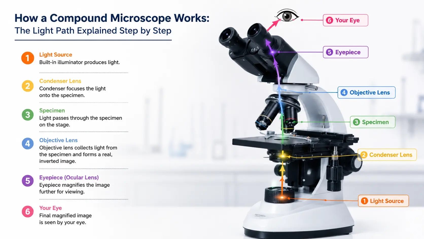
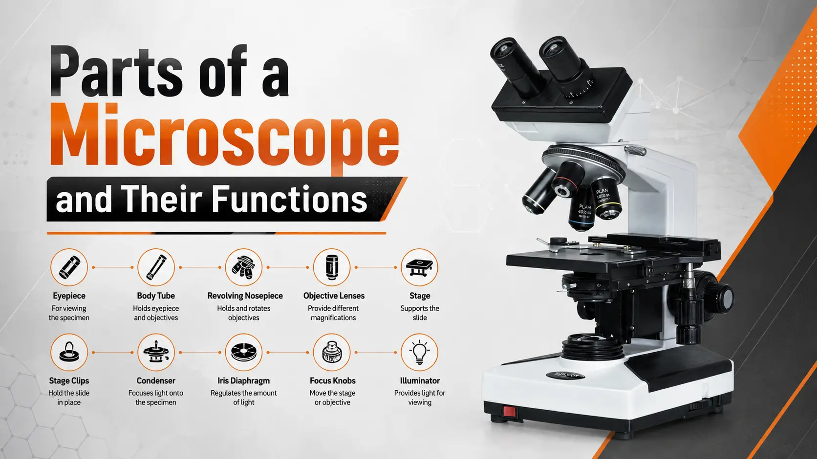
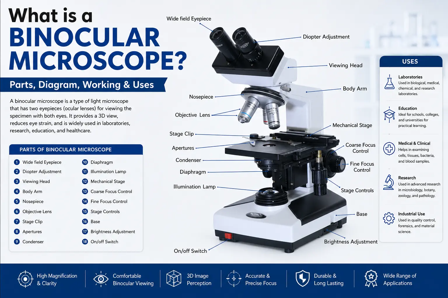
A compound microscope is an optical microscope that uses two lens systems — an objective lens near the specimen and an eyepiece near the eye — to magnify samples from 40x to 1000x. The objective forms a real inverted image, which the eyepiece magnifies again, giving high total magnification for cells and microorganisms.
This is the microscope most people picture when they hear the word.
Total magnification is simple to work out: multiply the eyepiece by the objective. A 10x eyepiece with a 40x objective gives 400x. Swap to the 100x oil immersion objective and you are at 1000x, which is about the practical ceiling for a light microscope because of the diffraction limit of visible light (roughly 200 nanometres).
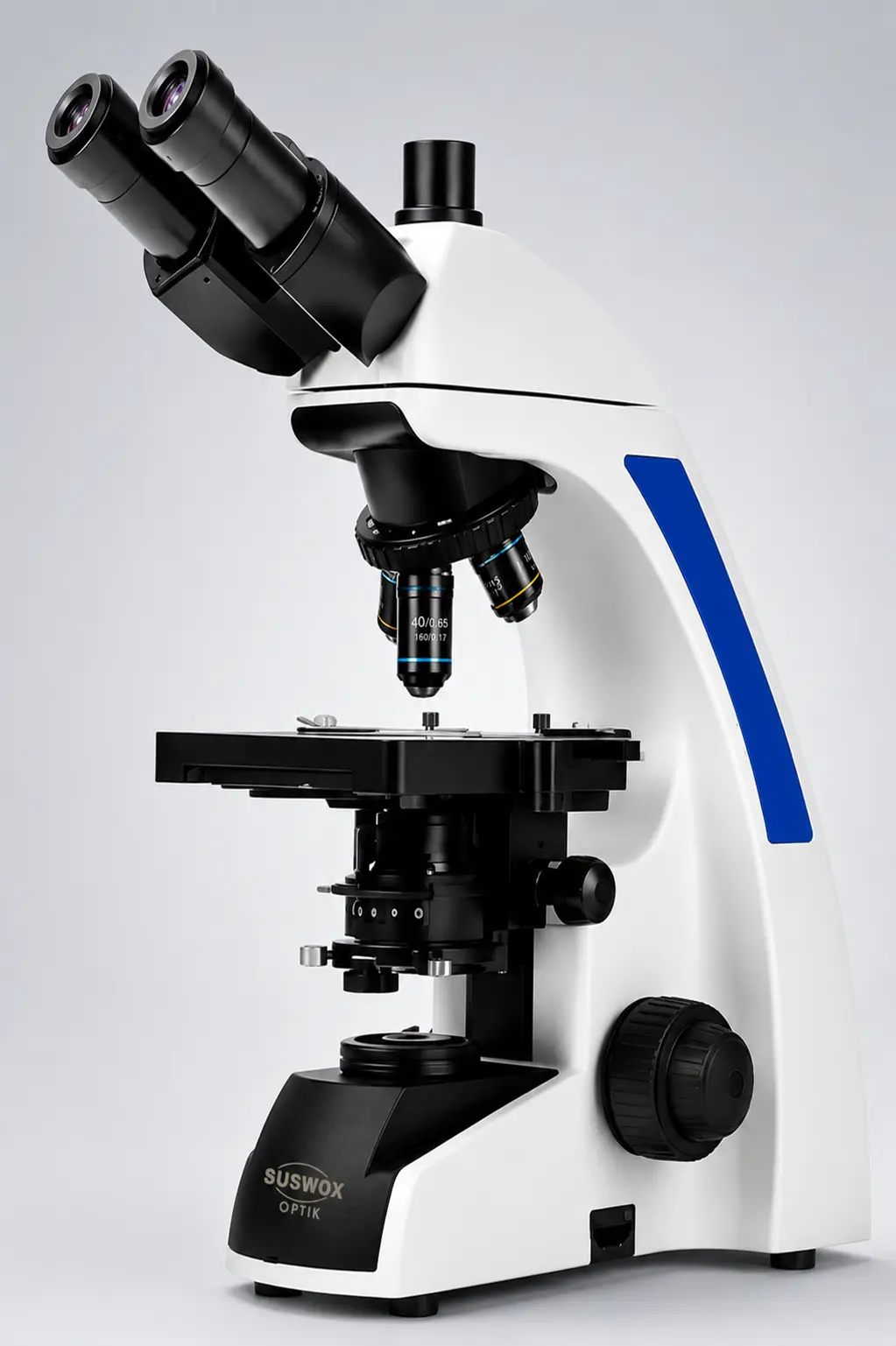
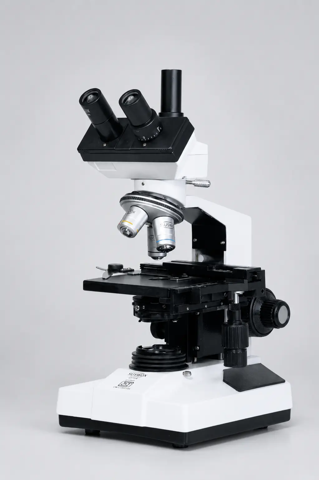
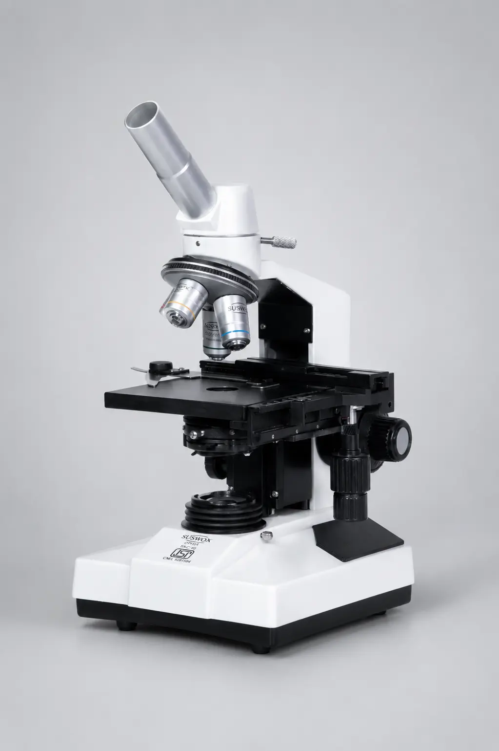
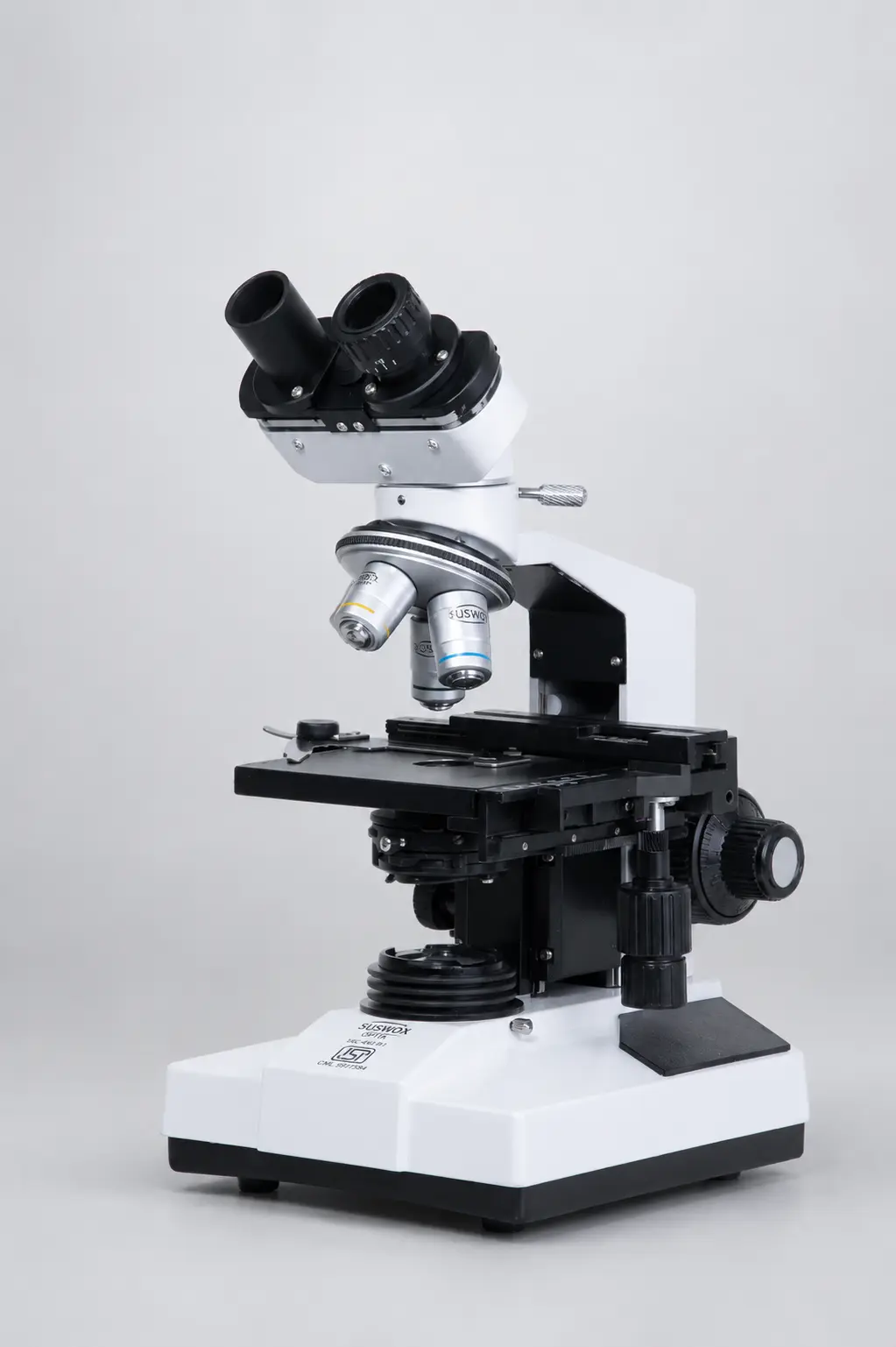
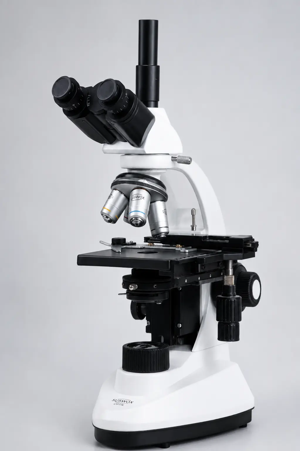
Compound microscopes come in three head styles, and this is where buyers usually get confused:
| Head Type | Eyepieces | Camera Port | Typical Use |
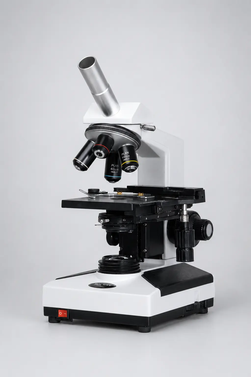
| Monocular | 1 | No | School labs, budget setups |
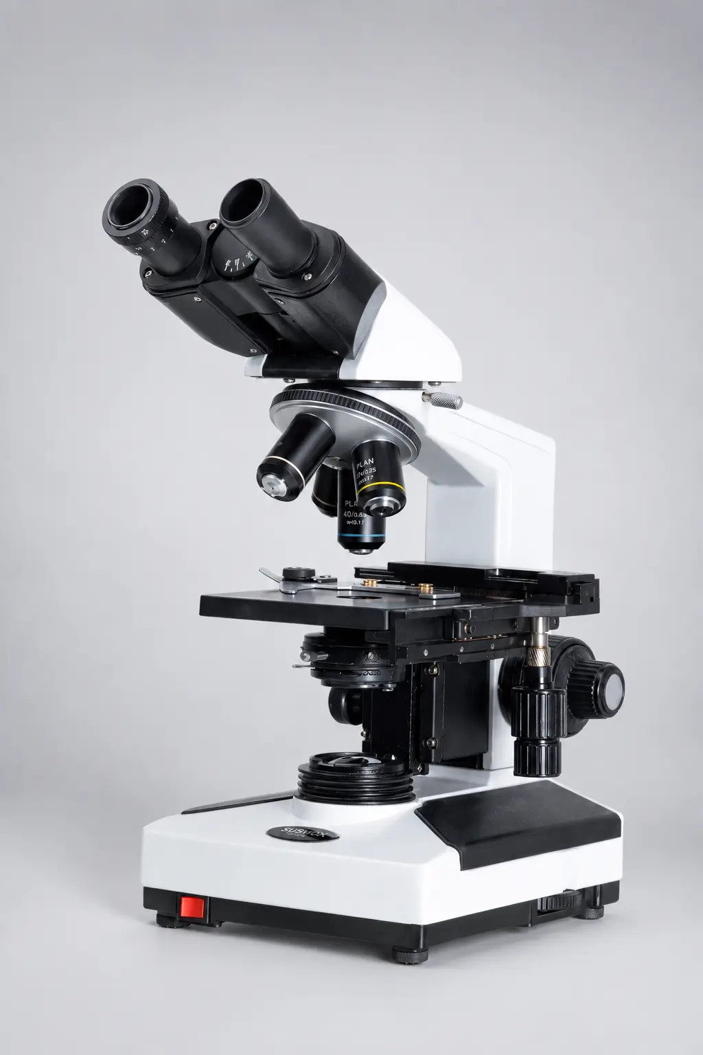
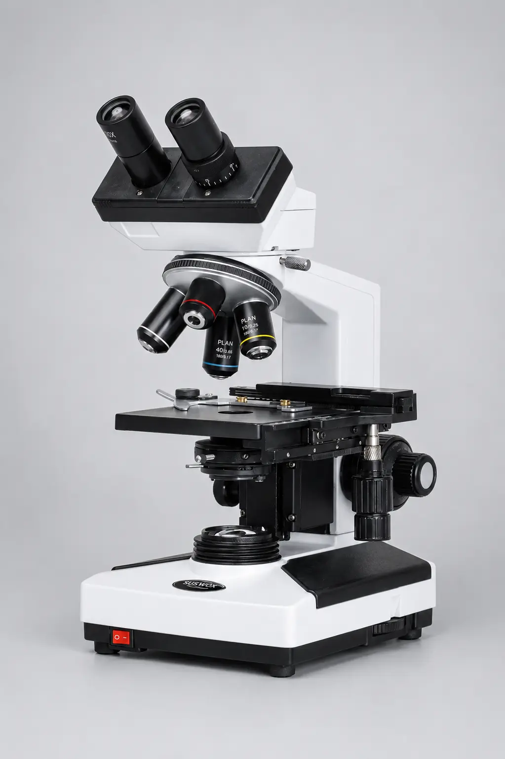
| Binocular | 2 | No | Pathology labs, daily diagnostic work |
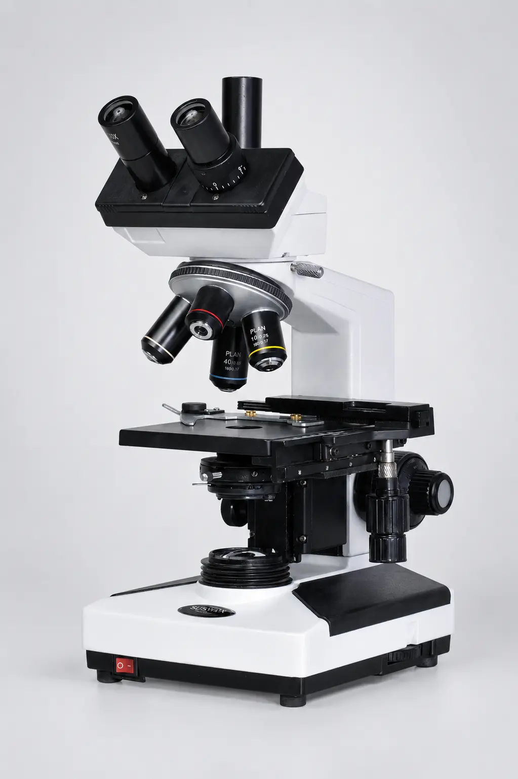
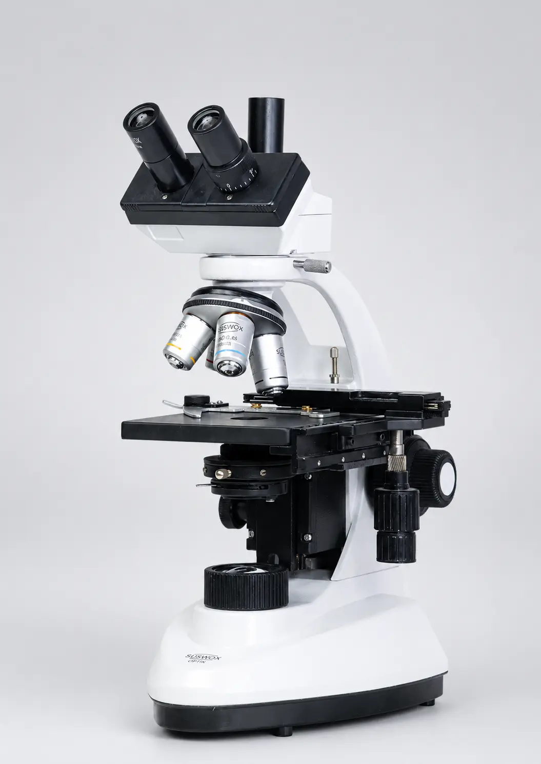
| Trinocular | 2 + 1 vertical tube | Yes | Documentation, teaching, publication imaging |
If technicians will sit at the scope for four hours a day, binocular is not a luxury. It is eye-strain prevention. We cover this in detail in our guide on what a binocular microscope is.
Where it is used:
- Pathology labs — blood smears, urine analysis, stool examination
- MBBS and BSc histology practicals
- Microbiology — bacterial staining and identification
- Water testing laboratories
- Veterinary diagnostics
Best for: any thin, transparent, stained specimen on a glass slide.
Suswox manufactures this category across its SC-300, SKC and CXL series lab microscopes in monocular, binocular and trinocular configurations.
[IMAGE 2 — file: compound-microscope-parts-labelled-diagram.webp | alt: “Labelled diagram of compound microscope showing eyepiece, objective lenses, stage and condenser”]
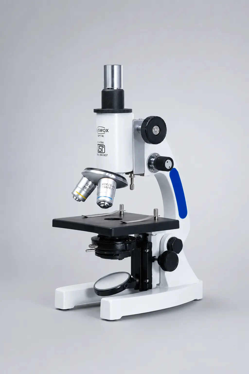
3. Student Microscope
Technically a student microscope is a compound microscope. But in Indian procurement it is treated as its own category, so it deserves its own entry.
The difference is build philosophy. A student microscope is designed to survive a school lab — where forty pairs of hands will touch it, the coarse focus will get yanked, and slides will get pushed onto the stage sideways. So you get a simplified 3-objective turret, a rigid metal body, LED illumination that runs on both mains and battery, and a lower price.
Typical specification for a school-grade unit:
| Feature | Standard Configuration |
| Objectives | 4x, 10x, 40x (achromatic) |
| Eyepiece | WF 10x / WF 15x |
| Magnification range | 40x – 600x |
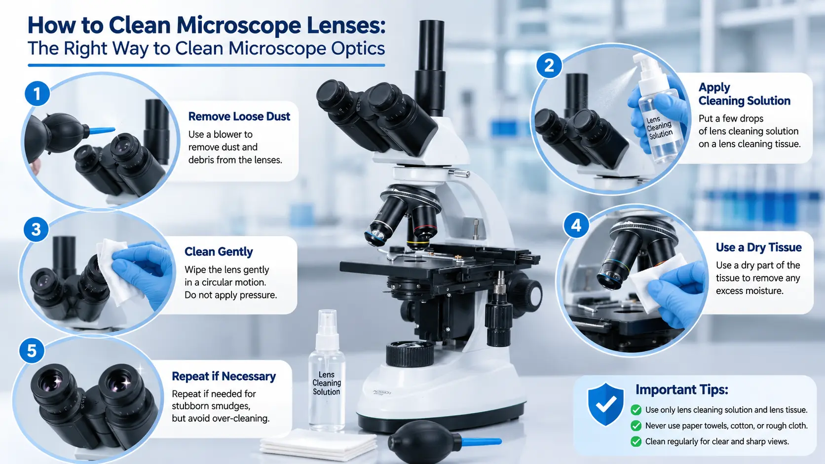
| Illumination | LED, rechargeable + mirror option |
| Stage | Plain with clips or mechanical |
| Body | Die-cast aluminium |
And this is the practical bit most purchase committees miss: rechargeable LED matters enormously in schools where power cuts are routine. A microscope that stops working at 11 am is a microscope that does not get used.
Where it is used:
- CBSE, ICSE and state board school biology labs
- BSc and BEd college practicals
- NEET coaching institute laboratories
- Science exhibitions and Atal Tinkering Labs
Best for: bulk institutional purchase where durability beats optical perfection.
Browse the student microscope range here — SK-33, SK-44, SK-66 and SK-555 models are built specifically for this use.
[IMAGE 3 — file: student-microscope-school-biology-lab.webp | alt: “Student microscope with LED illumination used in a school biology laboratory in India”]
4. Stereo Zoom Microscope
What is a stereo microscope used for?
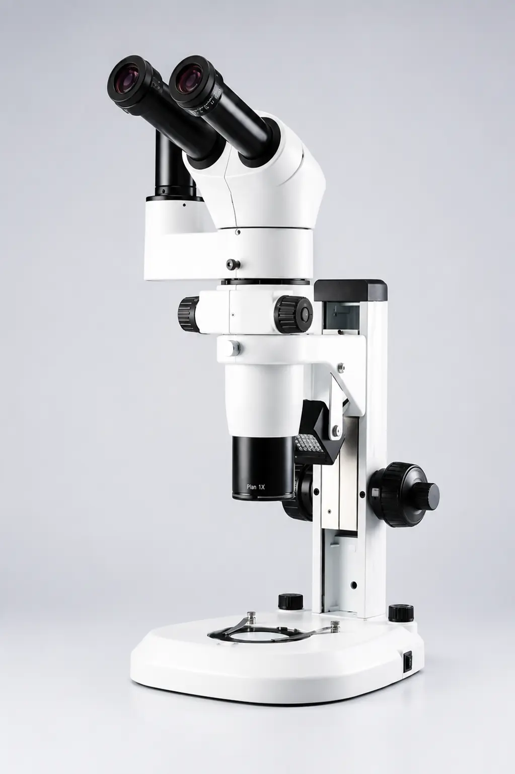
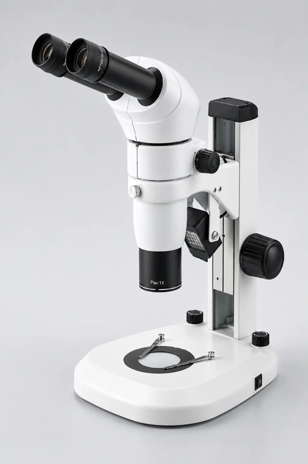
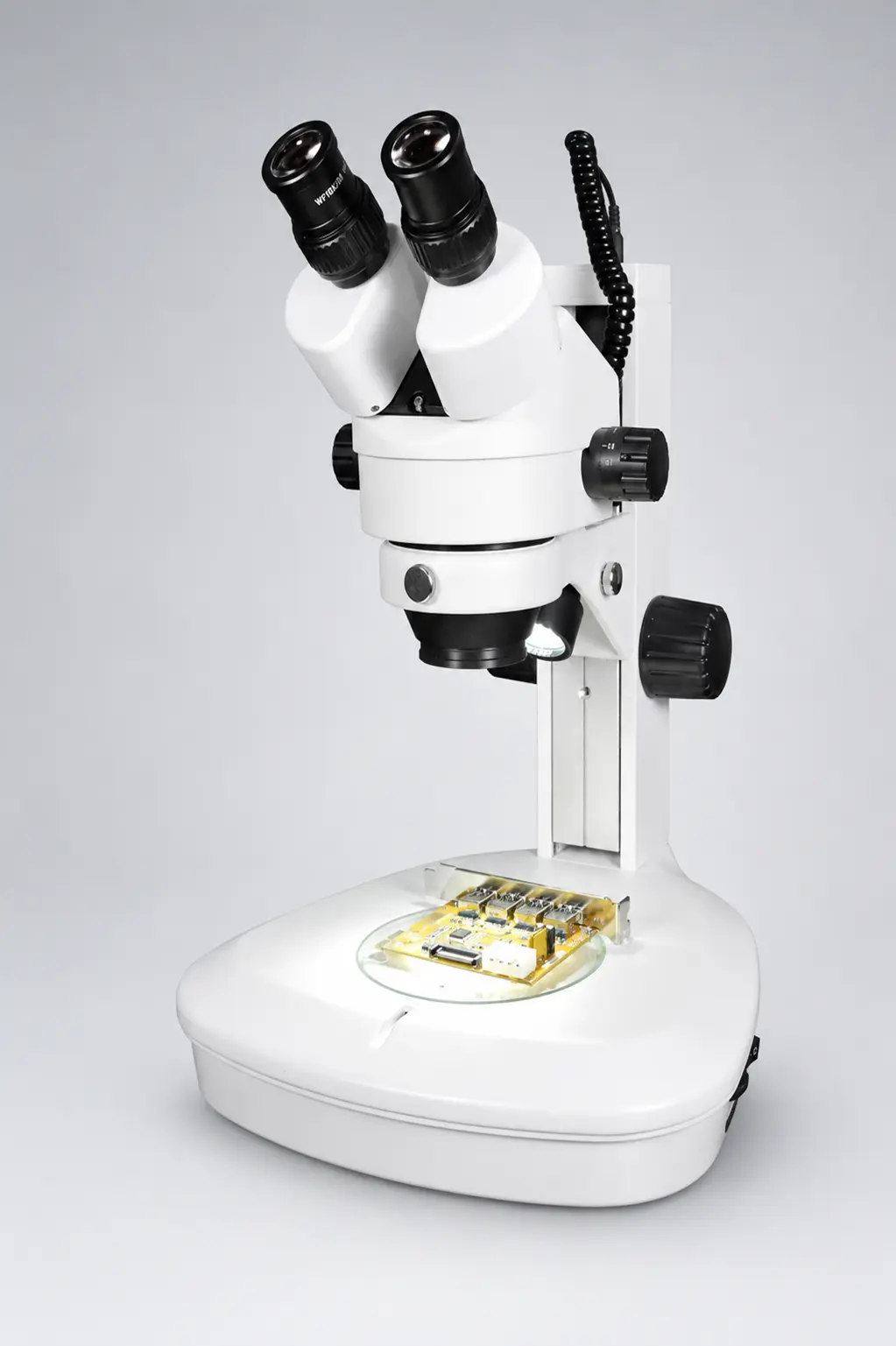
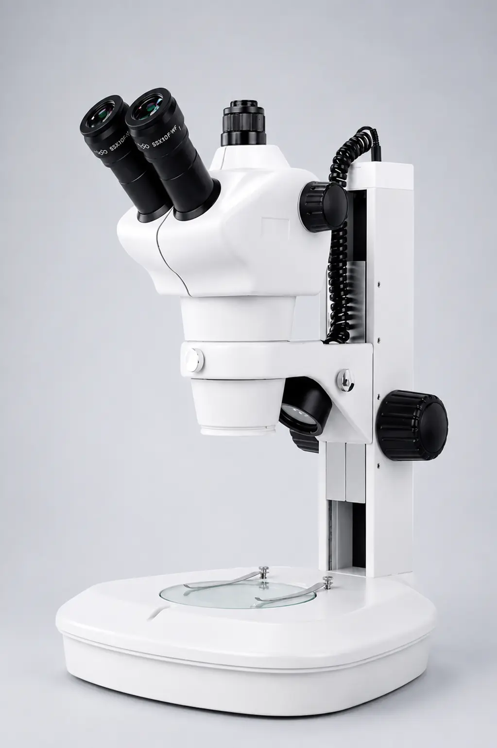
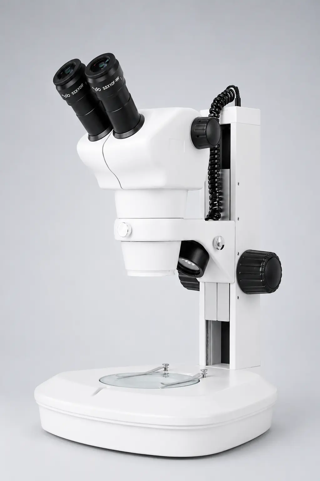
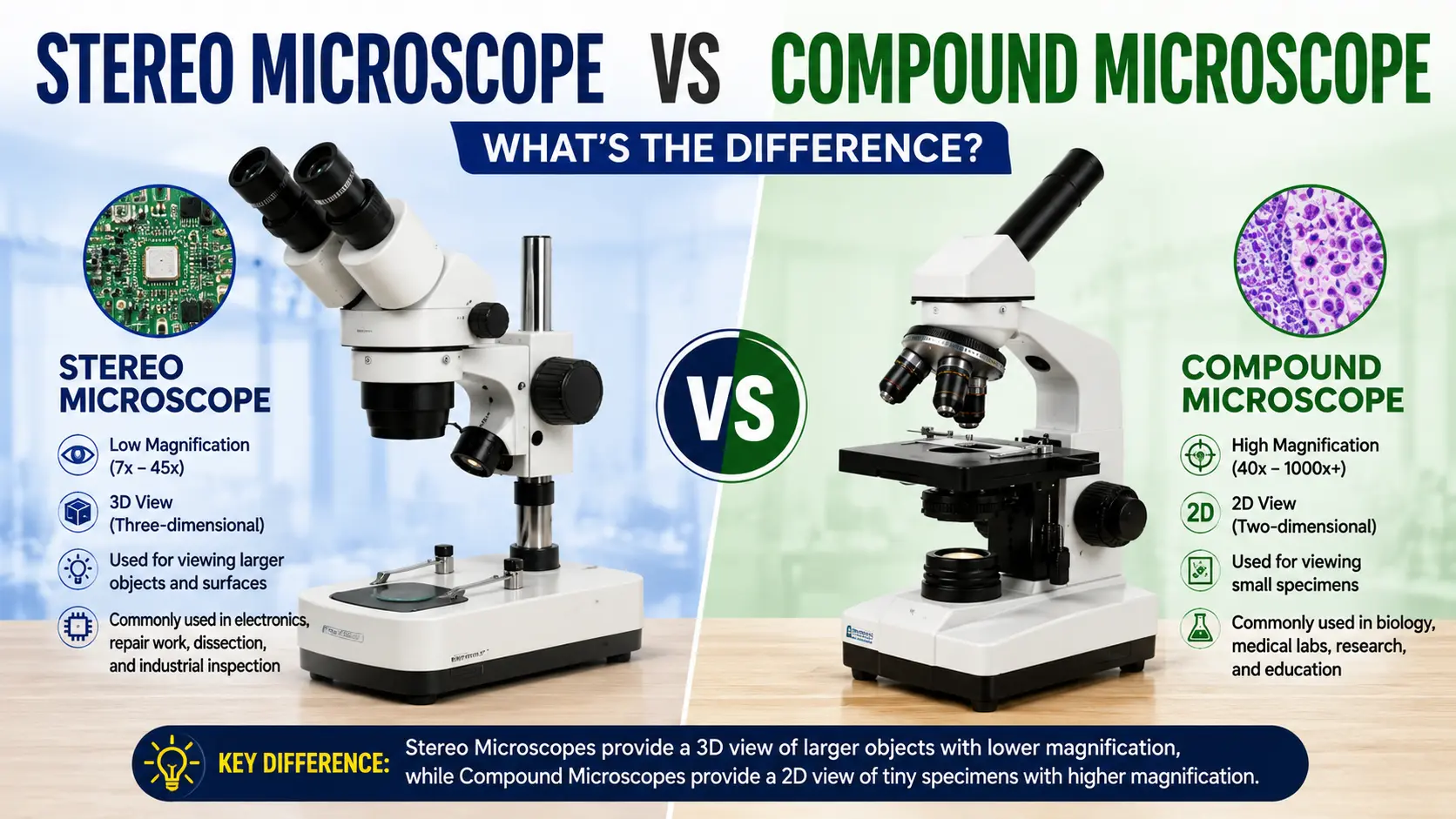
A stereo microscope, also called a dissecting microscope, uses two separate optical paths to give a three-dimensional, right-side-up view of solid objects at 6.5x to 90x magnification. It is used for dissection, PCB inspection, soldering, gem grading and any task where you need to work with your hands under the lens.
The 3D part is the whole point.
Under a compound microscope everything is flat and reversed — move the slide left, the image goes right. Try soldering a component while looking through that and you will destroy the board. A stereo microscope keeps depth and direction intact, and leaves 80–100 mm of working distance under the objective for tools.
Where it is used:
- Electronics — PCB rework, chip inspection, wire bonding
- Zoology and botany dissection practicals
- Gemmology and jewellery setting
- Watch and precision mechanical assembly
- Forensic examination of fibres, coins, tool marks
- Textile fibre and weave analysis
Best for: anything opaque, solid, and larger than a slide.
Suswox produces the STZ and SPZ series in trinocular and binocular heads — see the stereo zoom microscope category.
[IMAGE 4 — file: stereo-zoom-microscope-pcb-inspection.webp | alt: “Stereo zoom microscope used for PCB inspection and electronic component soldering”]
5. Inverted Microscope
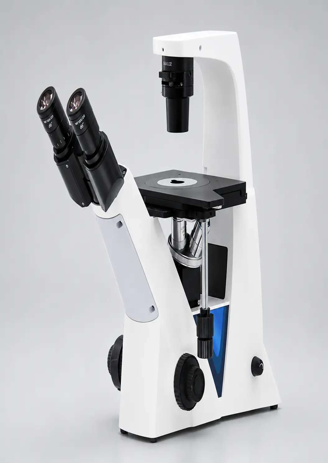
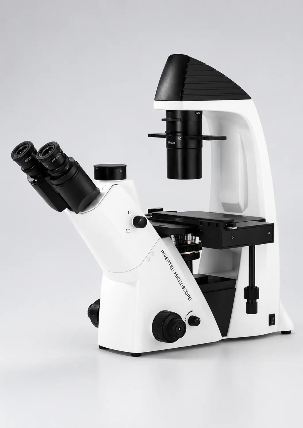
Turn a compound microscope upside down. Objectives below the stage, illumination above. That is an inverted microscope, and there is a very specific reason it exists.
Cells growing in a culture flask sit at the bottom of the flask, in liquid. You cannot get a conventional objective anywhere near them — the flask wall and the medium are in the way. Flip the optics underneath, and suddenly you can view living cells through the thin base of the vessel without opening it or contaminating it.
That single design change made modern cell biology possible.
Where it is used:
- Tissue culture and cell line maintenance
- IVF clinics — oocyte and embryo assessment
- Pharmaceutical cytotoxicity studies
- Metallurgy — examining large, heavy, flat metal samples
- Microbiology of live cultures in petri dishes
Best for: living cells in liquid, and heavy samples that cannot be inverted onto a stage.
Suswox manufactures the IMX-Ti in this category. Full details on the inverted microscope page.
[IMAGE 5 — file: inverted-microscope-tissue-culture-lab.webp | alt: “Inverted microscope viewing cell culture flask in a tissue culture laboratory”]
6. Digital Microscope
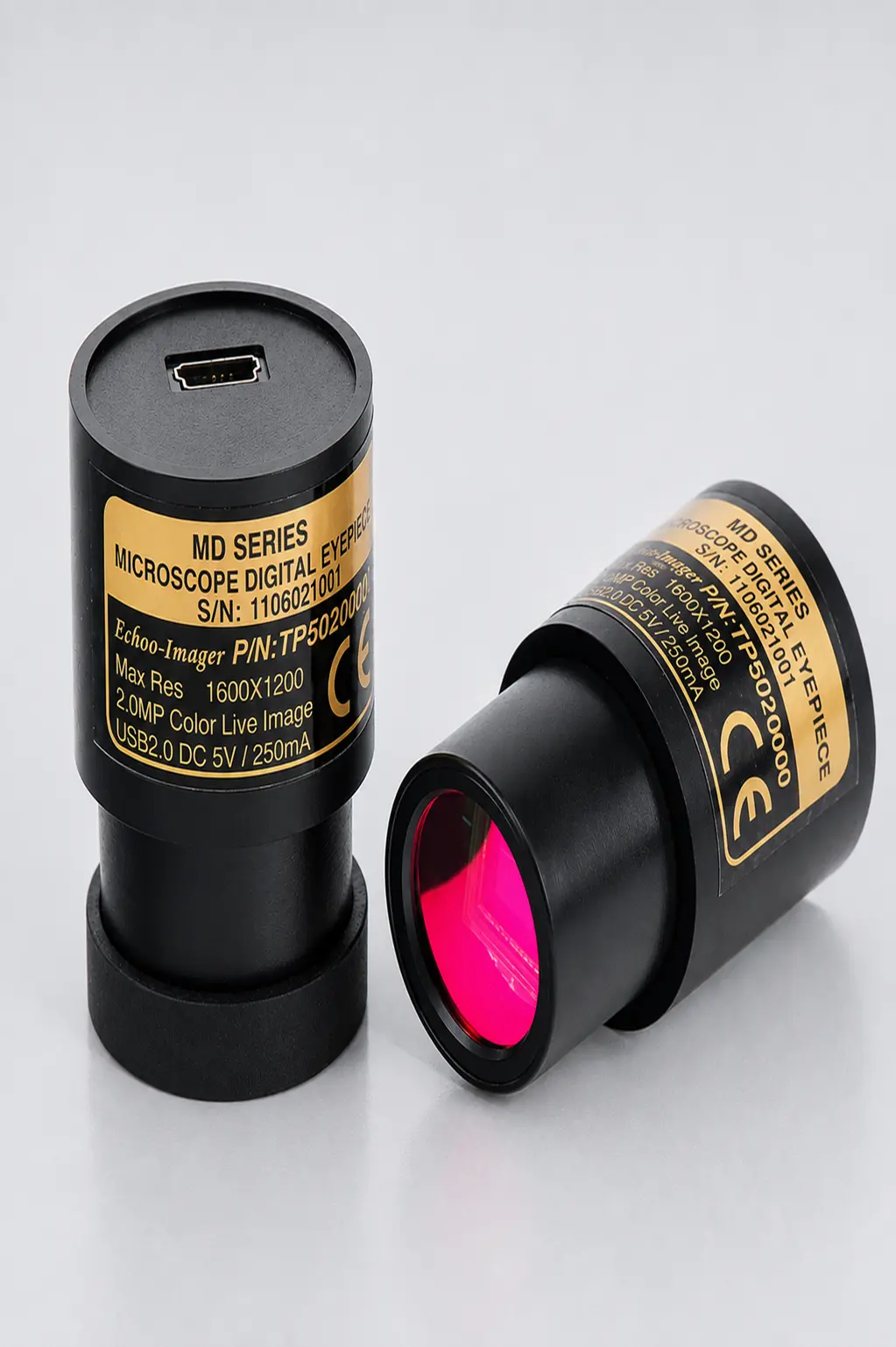
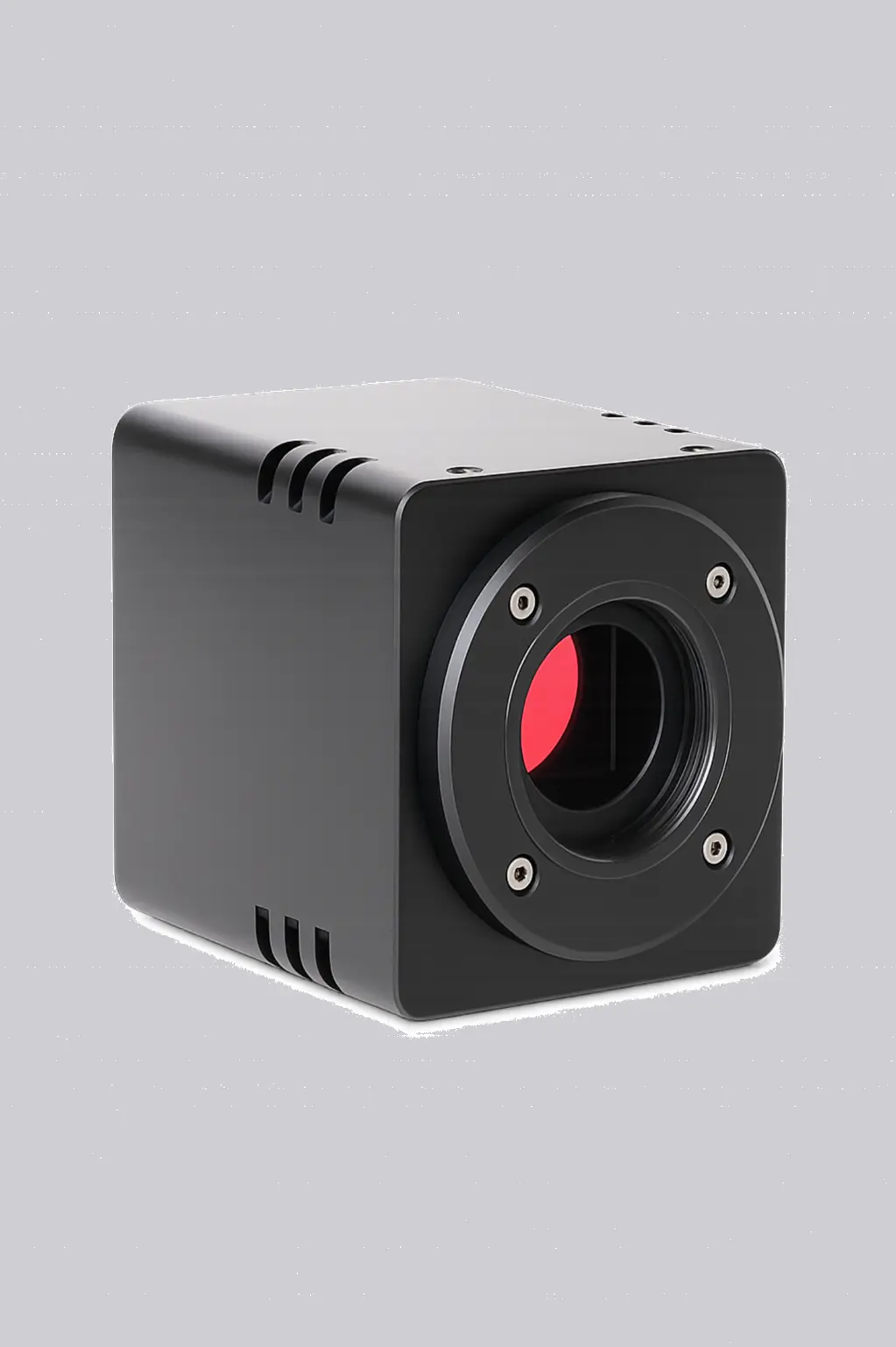
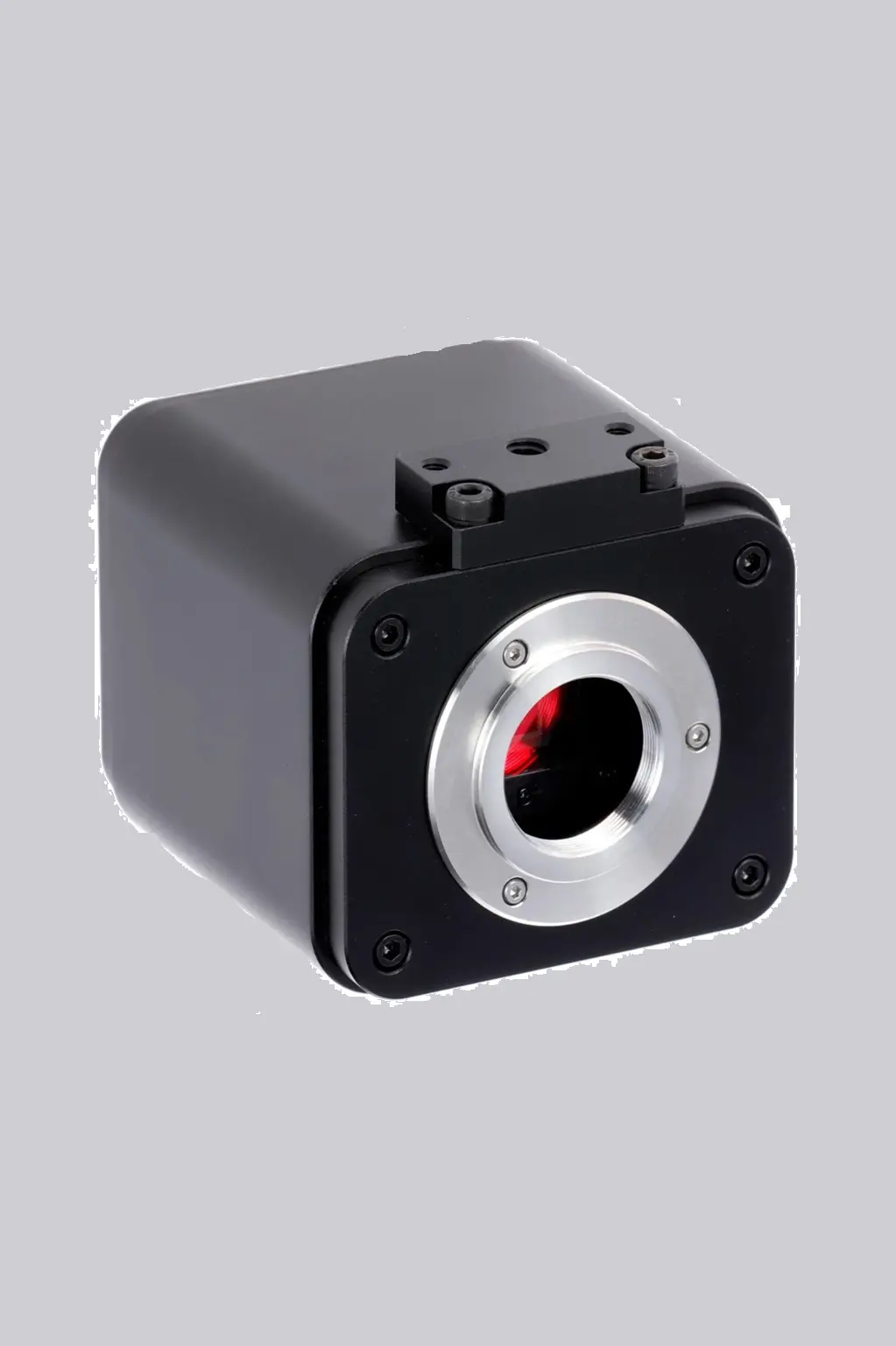
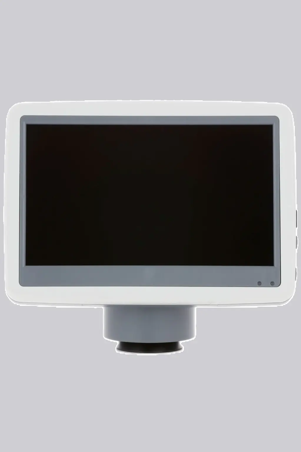
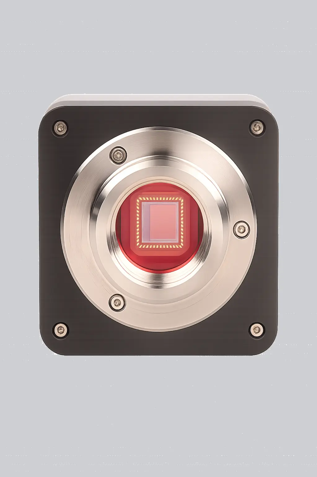
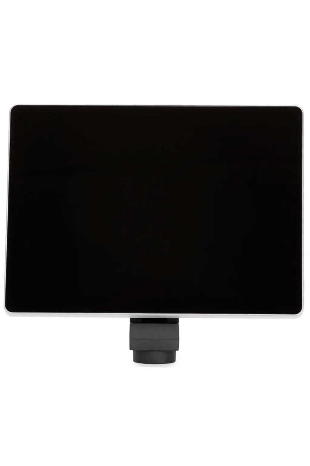
A digital microscope replaces the eyepiece with a camera sensor and sends the image to an LCD screen, monitor or computer. Some models keep the eyepieces and add a camera on the trinocular port; others drop eyepieces entirely.
The value is not really magnification. It is sharing.
When a professor is teaching thirty students, an eyepiece serves one person at a time. A digital microscope puts the same image on a screen for the whole room. When a QC inspector rejects a batch, a digital microscope produces a timestamped image file that goes into the report instead of a verbal description.
Two main formats:
| Format | Description | Suits |
| Camera + software | Camera mounts on a trinocular port, connects via USB or HDMI to a PC | Labs that already own good optics |
| Built-in LCD | Standalone unit with an integrated screen | Training rooms, industrial floor QC, field use |
Where it is used:
- Teaching and demonstration in colleges
- Industrial quality control with image records
- Mobile phone and micro-electronics repair
- Telepathology and second-opinion sharing
- Documentation for research papers and NABL audits
Best for: anywhere the image has to be seen by more than one person or saved as evidence.
Suswox offers the XCAM Mini, X Cam and AT-Series in this category — see digital microscopes.
[IMAGE 6 — file: digital-microscope-lcd-screen-camera.webp | alt: “Digital microscope with LCD display and camera used for on-screen sample viewing”]
7. Travelling Microscope
What is a travelling microscope used for?
A travelling microscope is a low-power compound microscope mounted on a vertical and horizontal vernier scale, used to measure very small distances that cannot be measured with a scale or vernier caliper. Its least count is typically 0.001 cm, and it is standard equipment in Class 11, Class 12 and BSc physics laboratories.
It does not really magnify for observation. It measures.
The least count is worked out the same way as any vernier instrument: divide the smallest main scale division by the number of vernier divisions. With a 0.5 mm main scale division and 50 vernier divisions, you get 0.5 ÷ 50 = 0.01 mm, which is 0.001 cm.
Standard physics experiments that need one:
- Refractive index of a glass slab by apparent depth method
- Radius of curvature of a lens surface
- Newton’s rings — measuring ring diameters
- Diameter of a capillary tube bore
- Surface tension by capillary rise method
Best for: physics practical labs and basic metrology work.
If your institution is stocking a physics lab, the travelling microscope range covers the standard SK-T2 configuration.
[IMAGE 7 — file: travelling-microscope-vernier-scale-physics-lab.webp | alt: “Travelling microscope with vernier scale used in physics laboratory for least count measurement”]
8. Projection Microscope
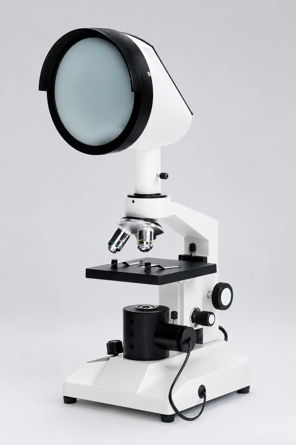
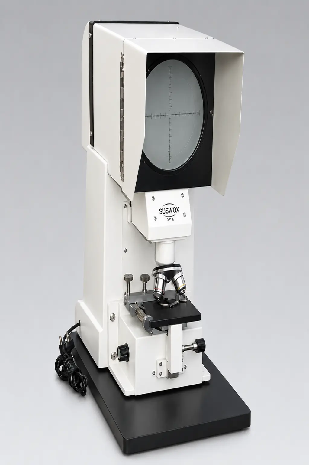
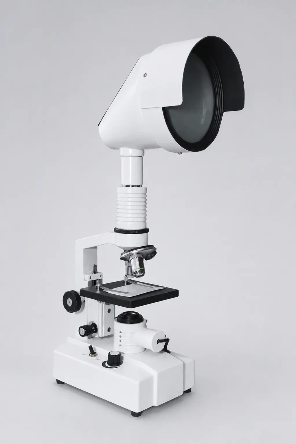
A projection microscope throws a magnified image of a slide onto a screen or wall so a whole class can see it at once. Older models used a strong lamp and a mirror system; newer ones combine optics with a digital projector.
Honestly, this category has shrunk since digital microscopes arrived. But it still has a place — mainly in schools that want a simple, no-computer, no-software way of showing a specimen to a full classroom.
Where it is used:
- Classroom biology demonstrations
- Science exhibitions and open days
- Museum and planetarium displays
- Group training sessions
Best for: teaching environments without IT infrastructure.
See the projection microscope models here.
[IMAGE 8 — file: projection-microscope-classroom-demonstration.webp | alt: “Projection microscope displaying a magnified slide image for classroom demonstration”]
9. Operating (Surgical) Microscope
An operating microscope is a stereo microscope built for the operation theatre. Long working distance, so the surgeon’s hands and instruments fit underneath. Coaxial illumination, so the light goes exactly where the surgeon is looking. Counterbalanced arm, so it stays where it is pushed.
Magnification usually stays modest — 4x to 40x. Surgeons do not need 1000x. They need a clear, bright, stable, three-dimensional view of tissue while both hands are busy.
Where it is used:
| Specialty | Typical Procedure |
| Ophthalmology | Cataract surgery, corneal transplant |
| ENT | Middle ear surgery, tympanoplasty |
| Dentistry | Root canal treatment, endodontics |
| Neurosurgery | Microvascular procedures |
| Plastic surgery | Microvascular anastomosis, replantation |
Best for: clinical procedures needing magnified vision and free hands.
Suswox supplies this category through its operating microscope range, including units in use at government medical colleges.
[IMAGE 9 — file: operating-microscope-surgical-ent-dental.webp | alt: “Operating microscope used in ENT and dental surgery with coaxial illumination”]
10. Polarizing Microscope
A polarizing microscope adds two polarizing filters — a polarizer below the stage and an analyser above it — plus a rotating stage. When light passes through a birefringent material between crossed filters, the sample lights up in vivid interference colours against a black background.
Those colours are data, not decoration. They tell a geologist which mineral is in the rock and a pharmacist whether a drug has crystallised in the right polymorphic form.
Where it is used:
- Geology and mineralogy — thin section petrography
- Pharmaceutical crystallography and polymorph screening
- Polymer and fibre orientation studies
- Asbestos identification
- Clinical gout diagnosis — identifying urate crystals in joint fluid
Best for: materials whose internal crystal structure matters.
11. Fluorescence Microscope
A fluorescence microscope illuminates a specimen with a specific wavelength — usually blue, UV or LED-generated — which excites fluorescent dyes in the sample. Those dyes then emit light at a longer wavelength, and filters block everything except that emitted light. The result is a bright structure glowing on a black background.
In India, its most important everyday role is tuberculosis diagnosis. Auramine-stained sputum smears read under LED fluorescence are read faster and with better sensitivity than conventional Ziehl-Neelsen microscopy, which is why the National TB Elimination Programme pushed LED fluorescence microscopes into district labs.
Where it is used:
- TB diagnosis in DMC and district laboratories
- Immunofluorescence assays — ANA, ANCA testing
- Malaria and parasitology
- Cancer research, live-cell imaging
- Botany — chlorophyll autofluorescence studies
Best for: detecting specific tagged molecules, not general morphology.
[IMAGE 10 — file: fluorescence-microscope-led-tb-diagnosis.webp | alt: “LED fluorescence microscope used for TB sputum smear diagnosis in a laboratory”]
12. Electron Microscope (SEM & TEM)
Light has a limit. Around 200 nanometres, visible light simply cannot resolve two points as separate — the Abbe diffraction limit. To go beyond that you need something with a much shorter wavelength, and Ernst Ruska’s answer in 1931 was to use a beam of electrons focused by electromagnetic lenses. He received the Nobel Prize in Physics for it in 1986.
There are two main types:
| Feature | SEM (Scanning) | TEM (Transmission) |
| How it works | Scans the surface with a beam | Passes the beam through the sample |
| Image | 3D surface topography | 2D internal structure |
| Magnification | Up to ~1,00,000x | Up to ~10,00,000x |
| Sample prep | Gold/carbon coating, dried | Ultra-thin sections (<100 nm) |
| Typical use | Fractures, powders, nanoparticles | Viruses, cell organelles, crystal lattice |
Where it is used:
- Nanotechnology and materials science research
- Virology — this is how viruses are actually visualised
- Semiconductor failure analysis
- Forensic examination of gunshot residue
- Metallurgical fracture analysis
Best for: anything below 200 nm.
Reality check: these are institutional instruments. They need a vacuum system, vibration isolation, a dedicated room and an operator. Most Indian users access them through central instrumentation facilities at IITs, CSIR labs or state universities rather than buying one.
[IMAGE 11 — file: scanning-electron-microscope-sem-lab.webp | alt: “Scanning electron microscope in a materials research laboratory”]
Optical vs Electron Microscope: What’s the Difference?
An optical microscope uses visible light and glass lenses to magnify samples up to about 1000x, and can view living specimens in colour. An electron microscope uses a focused electron beam and magnetic lenses to reach over 1,00,000x, giving far higher resolution, but samples must be dead, dried and placed in a vacuum.
| Point | Optical Microscope | Electron Microscope |
| Source | Visible light (LED/halogen) | Electron beam |
| Lenses | Glass | Electromagnetic |
| Max useful magnification | ~1000x – 1500x | 1,00,000x – 10,00,000x |
| Resolution limit | ~200 nm | ~0.1 nm |
| Living samples | Yes | No |
| Colour | Yes | Greyscale (false colour added later) |
| Environment | Normal room | Vacuum chamber |
| Cost in India | ₹3,000 – ₹12 lakh | ₹40 lakh and above |
| Operator training | Minimal | Specialised |
Worth knowing: the 200 nm barrier is not absolute any more. The 2014 Nobel Prize in Chemistry went to Eric Betzig, Stefan Hell and William Moerner for super-resolved fluorescence microscopy, which pushes light microscopy into the nanoscale. But those systems are research-grade and priced accordingly.
How to Choose the Right Type of Microscope
Start with the sample, not the specification sheet. Almost every wrong purchase we see started with someone comparing magnification numbers instead of asking what they were going to look at.
Four questions settle it:
1. Is your sample transparent or opaque? Transparent and mounted on a slide → compound microscope. Opaque and solid → stereo microscope. This single question eliminates half the catalogue.
2. Do you need to work with your hands under the lens? Dissection, soldering, repair → stereo, every time. The working distance and 3D view are non-negotiable.
3. Does the image need to leave the room? Reports, teaching, audits, second opinions → trinocular head with a camera, or a digital model.
4. How many hours a day? Under an hour → monocular is fine. More than two hours → binocular, and pay attention to interpupillary adjustment and diopter correction. Eye fatigue is a real operational cost in diagnostic labs.
One more thing that matters specifically in India: fungus. Humid coastal and monsoon conditions grow fungus on internal lens surfaces, and a fungal bloom on an objective is expensive to fix. Ask whether the optics are anti-fungal treated before you buy. Then store the instrument with silica gel under a dust cover.
For a deeper walkthrough, read our guide on how to choose the right microscope for your research.
Buying Guide: Microscope Manufacturer in India
Here is the part most listicles skip.
Almost every serious optical instrument in India traces back to a handful of manufacturing clusters, and Ambala in Haryana is the biggest of them — the town has been making scientific and optical instruments since before independence, and supplies laboratories across the country and overseas.
Suswox (Sudheer Scientific Works) is a microscope manufacturer in India based at 1265 Bengali Mohalla, Ambala, Haryana, manufacturing optical microscopes since 1973. The company produces student, laboratory, stereo zoom, inverted, digital, travelling, projection and operating microscopes, and supplies institutions including AIIMS Bathinda, AIIMS Nagpur, AIIMS Bilaspur, BHU, Kurukshetra University, MDU Rohtak, Haffkine Institute and the Government of Himachal Pradesh.
Why this matters when you are comparing microscope suppliers in India:
- Direct from factory — no trading margin stacked on top of the instrument
- 50+ years of manufacturing, not rebadged imports
- Spare parts and service available for the life of the instrument
- GeM and tender documentation support for government purchase
- Custom configurations — objectives, illumination and heads specified to your requirement
- Bulk institutional supply for schools, colleges and hospital labs
That last point separates real microscope manufacturers from resellers. When a district hospital needs eleven identical binocular microscopes with matching optics and a common service contract, a trading company cannot deliver that consistently. A factory can.
We have also listed the wider industry landscape in our post on the top 10 microscope manufacturers in India.
Get a quote: Suswox — 1265 Bengali Mohalla, Ambala (HR), India 133001 Phone: +91 8727015757 | +91 9416025615 Email: sales@suswox.com Browse all microscopes | Request a quote
Frequently Asked Questions
How many types of microscopes are there?
There are 12 commonly used types of microscopes: simple, compound, student, stereo zoom, inverted, digital, travelling, projection, operating, polarizing, fluorescence and electron microscopes. They fall into two families — optical microscopes that use light, and electron microscopes that use an electron beam.
Which type of microscope is used in schools?
Schools use student microscopes, which are compound microscopes with a simplified 3-objective turret, LED illumination and a rugged metal body. They magnify from 40x to about 600x, which covers every CBSE, ICSE and state board biology practical from onion peel cells to blood smears.
What is the difference between a compound and a stereo microscope?
A compound microscope gives a flat, inverted, high-magnification image of thin transparent samples on a slide. A stereo microscope gives a three-dimensional, upright, low-magnification image of solid objects, with enough working distance to use tools underneath. Compound for cells; stereo for circuit boards and dissection.
Which microscope has the highest magnification?
The transmission electron microscope (TEM) has the highest magnification, reaching roughly 10,00,000x with a resolution near 0.1 nanometre. Among optical microscopes, the compound microscope goes highest at about 1000x to 1500x using a 100x oil immersion objective.
What is the price of a microscope in India?
Microscope prices in India start around ₹3,000 for a basic student model and go up to ₹1,50,000 for a research-grade trinocular compound microscope. Stereo zoom microscopes run ₹12,000 to ₹80,000, inverted microscopes ₹60,000 upwards, and electron microscopes cost ₹40 lakh and above.
Can a microscope be used to see viruses?
No optical microscope can resolve most viruses, because they are smaller than the 200 nanometre diffraction limit of visible light. Viruses are visualised with transmission electron microscopes. Bacteria, however, are easily visible at 1000x on a standard compound microscope with oil immersion.
What is the least count of a travelling microscope?
The least count of a standard travelling microscope is 0.001 cm, or 0.01 mm. It is calculated by dividing the smallest main scale division (0.5 mm) by the number of vernier scale divisions (50), which gives 0.01 mm.
Which microscope is best for a pathology lab?
A binocular or trinocular compound microscope with 4x, 10x, 40x and 100x oil immersion achromatic objectives, LED Koehler illumination and an Abbe condenser is the standard choice for pathology labs. Trinocular is preferred where images must be recorded for reports or NABL documentation.