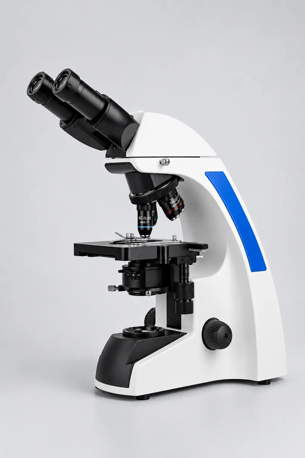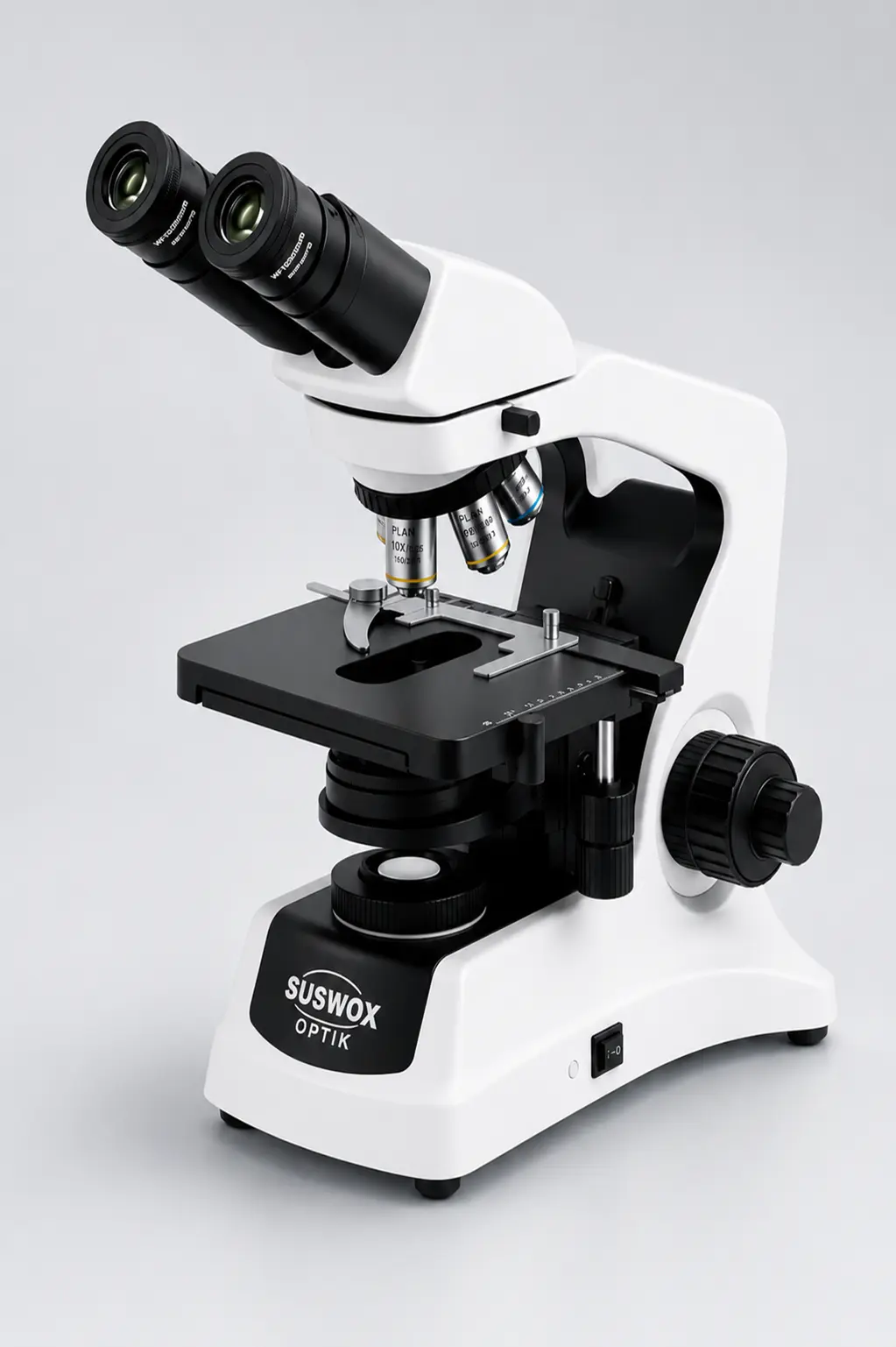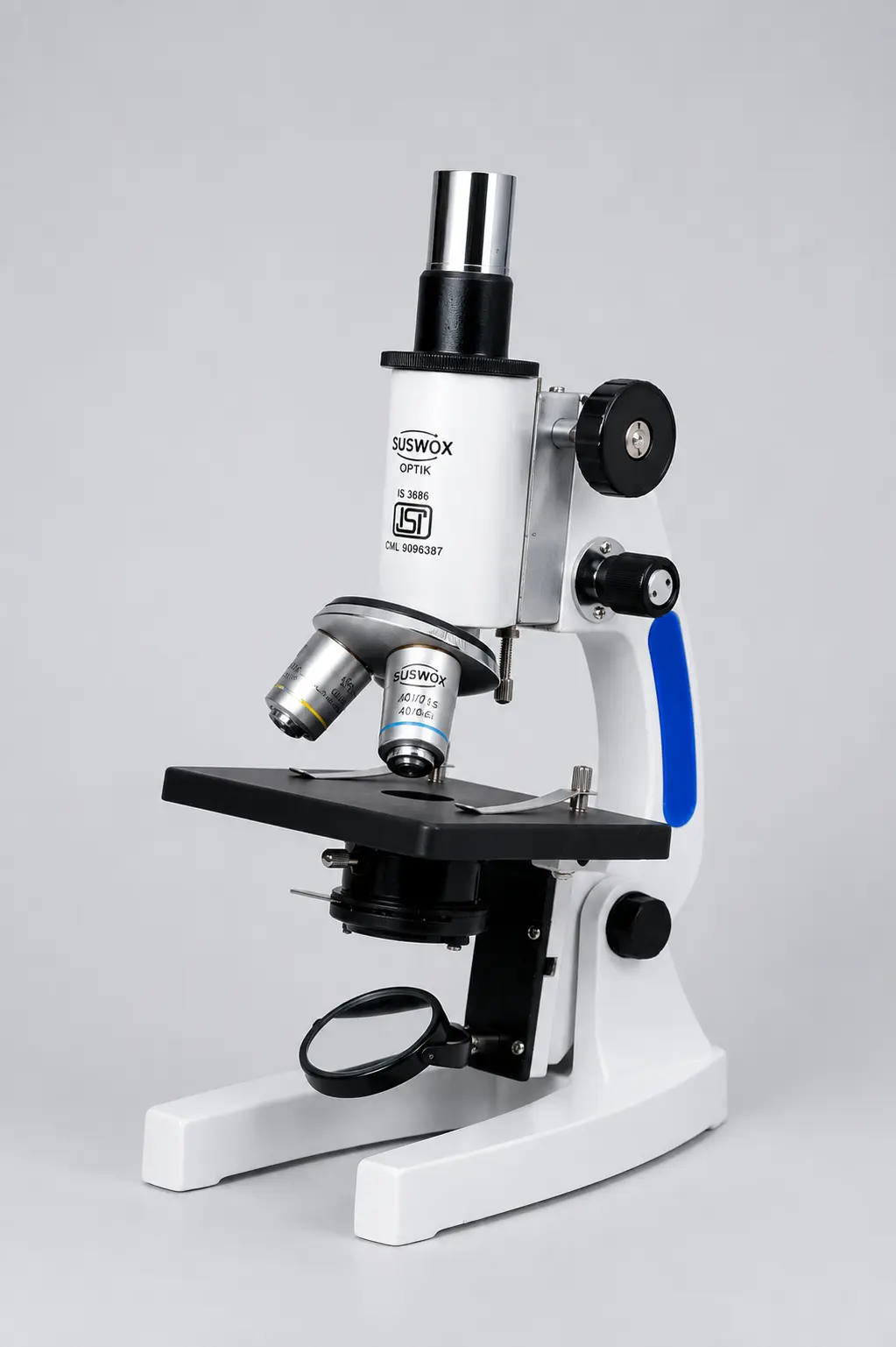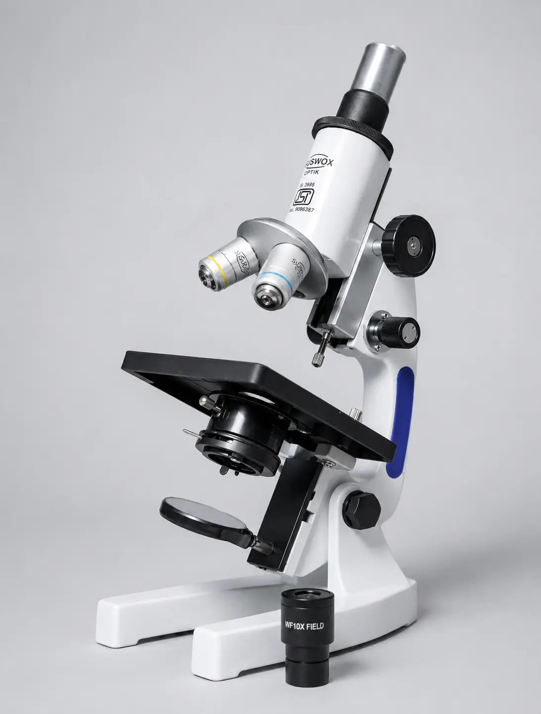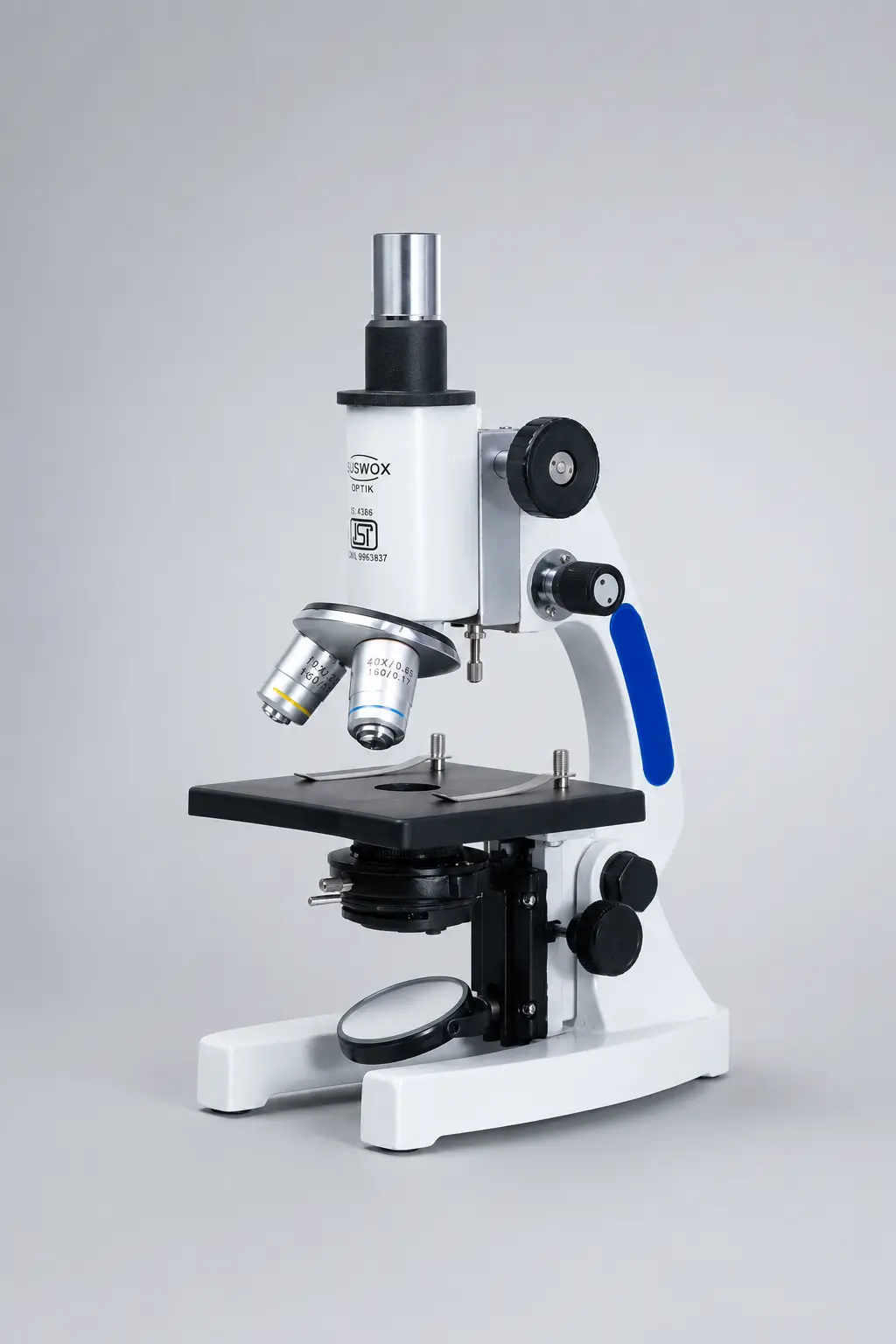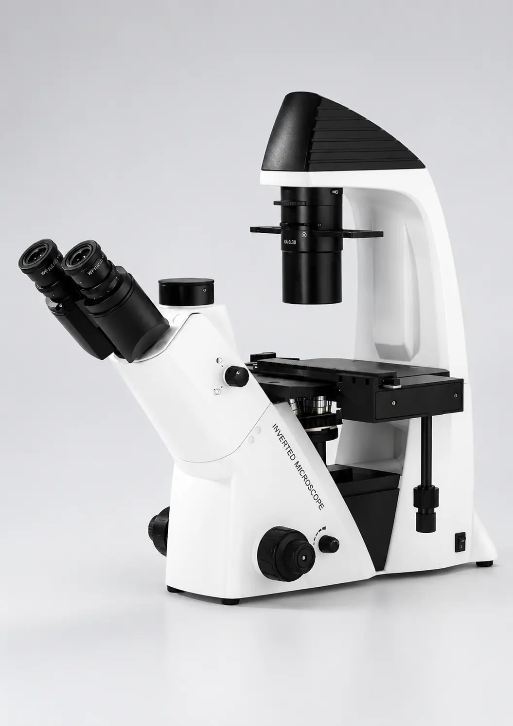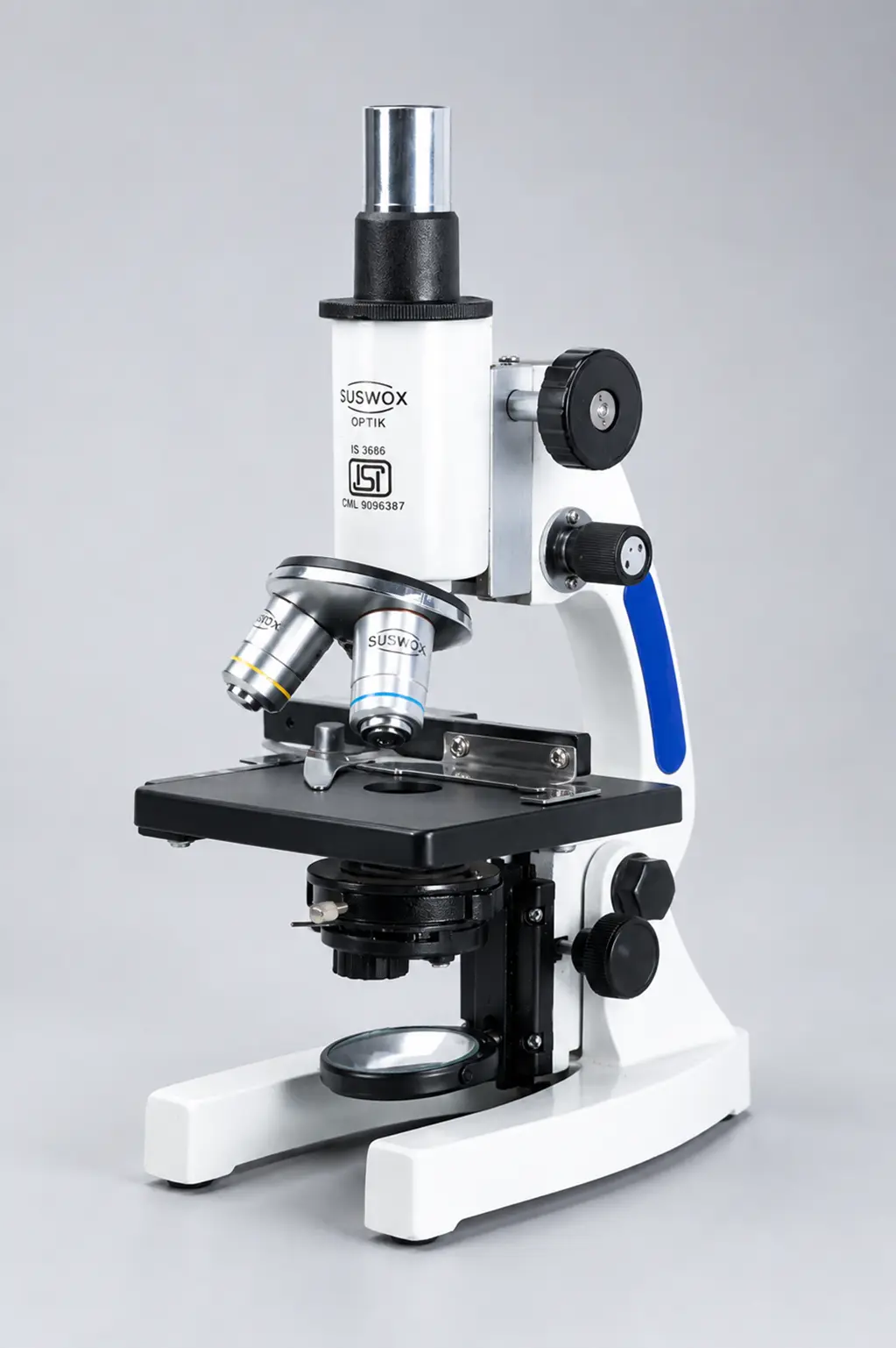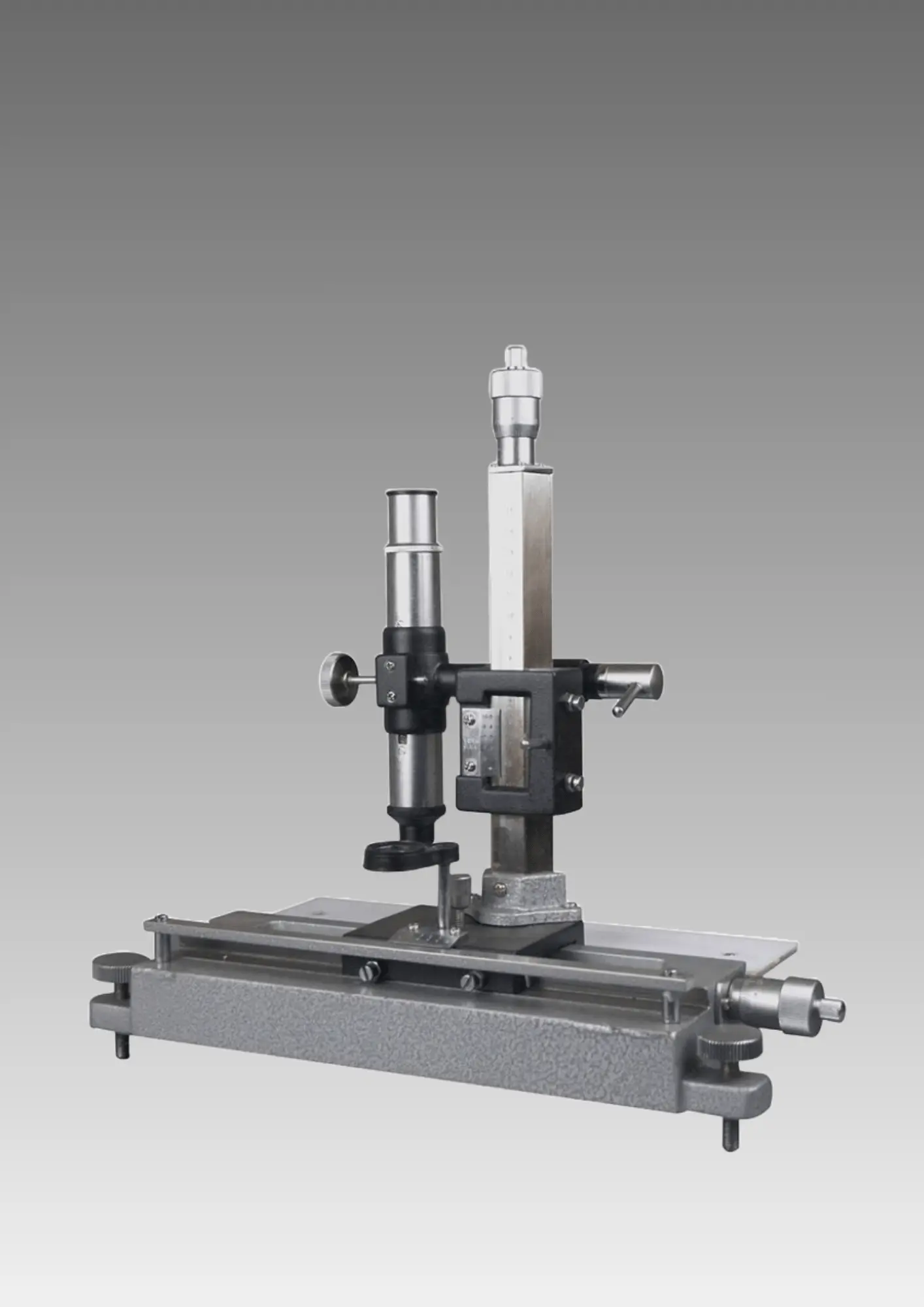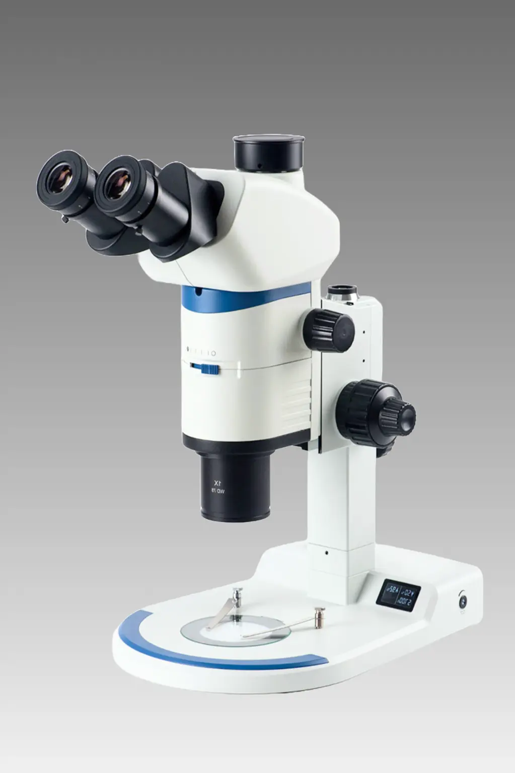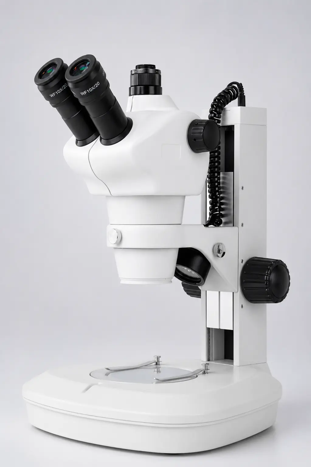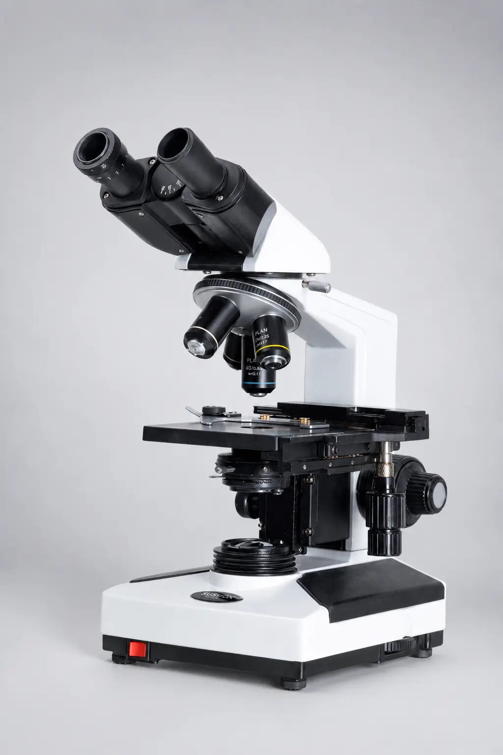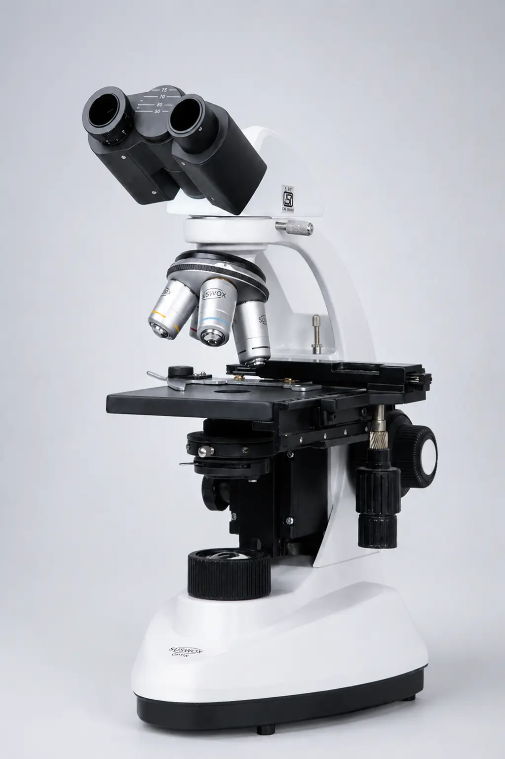
Picture a student leaning over a microscope for the very first time. One eye squeezed shut, the other watering after ten minutes, neck bent at a strange angle. Now picture the same student on a binocular microscope — both eyes open, relaxed, seeing the specimen in soft 3D. Same slide. Completely different experience.
That one change is the whole idea behind this instrument.
What is a binocular microscope?
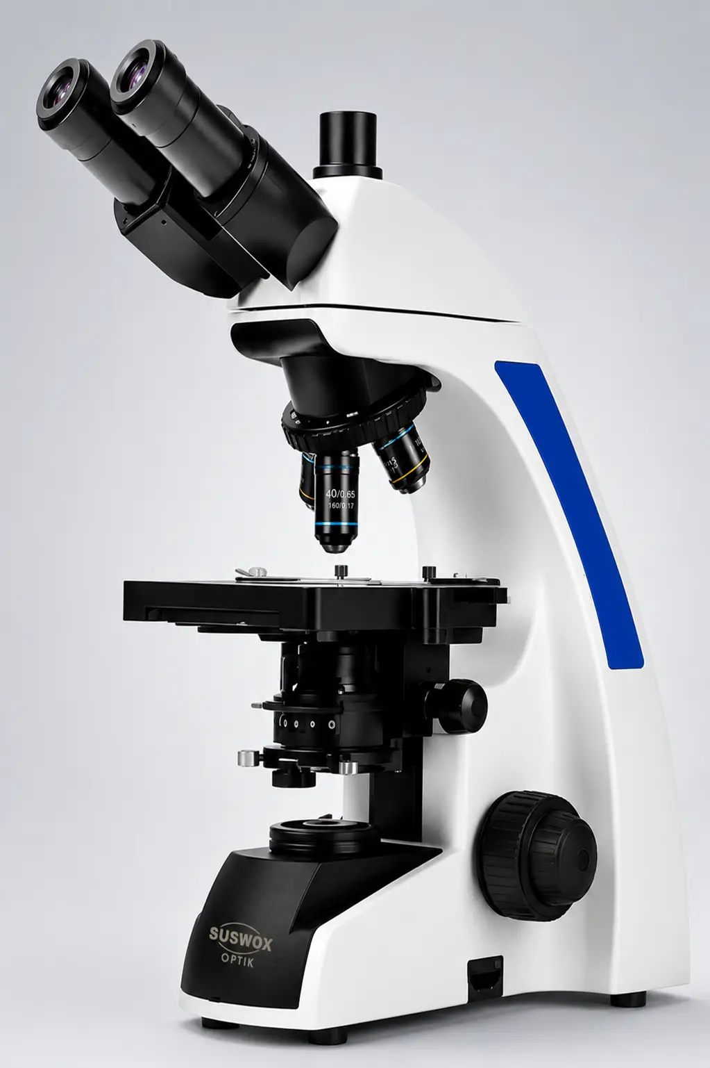
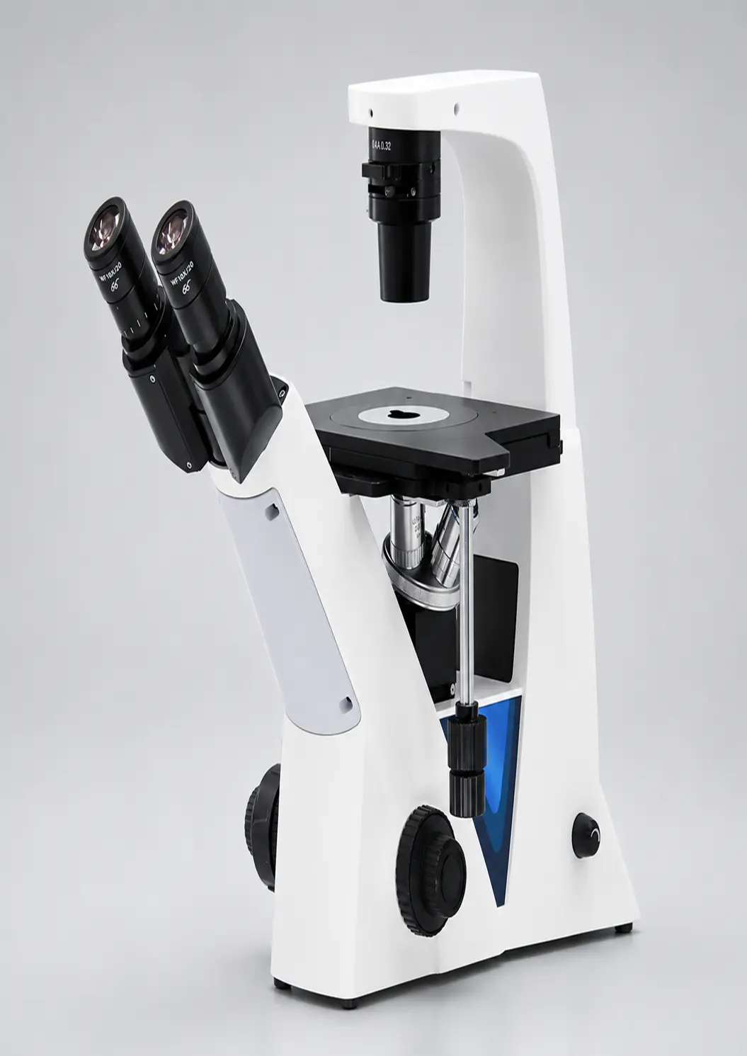
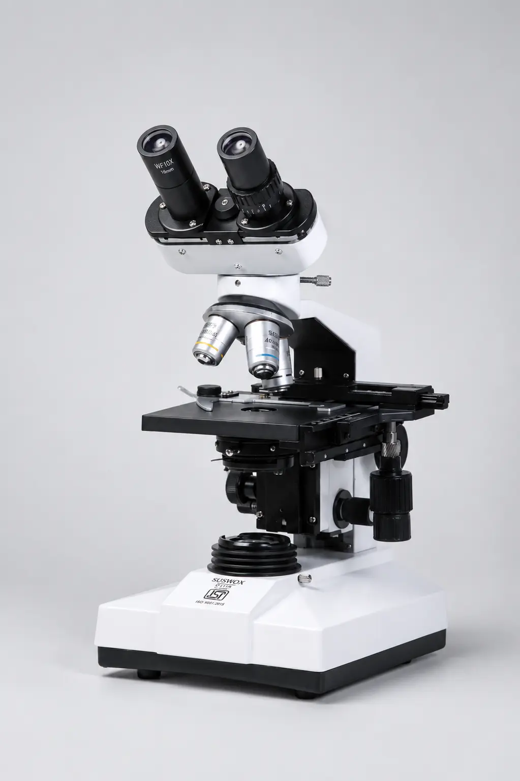
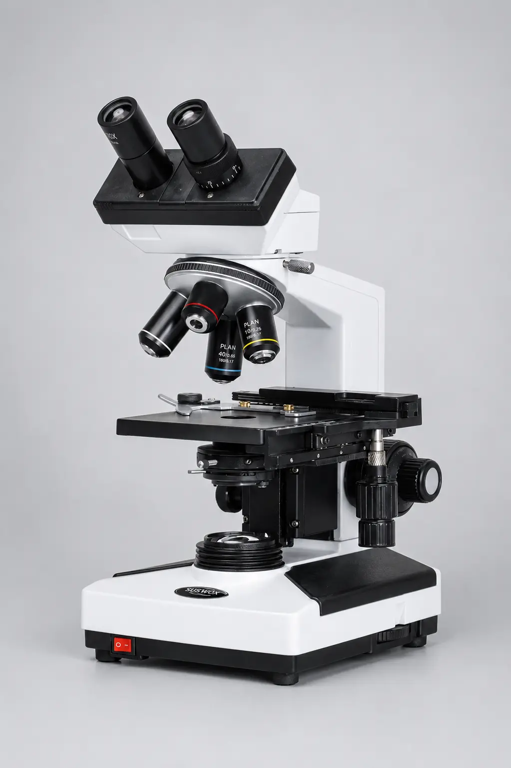
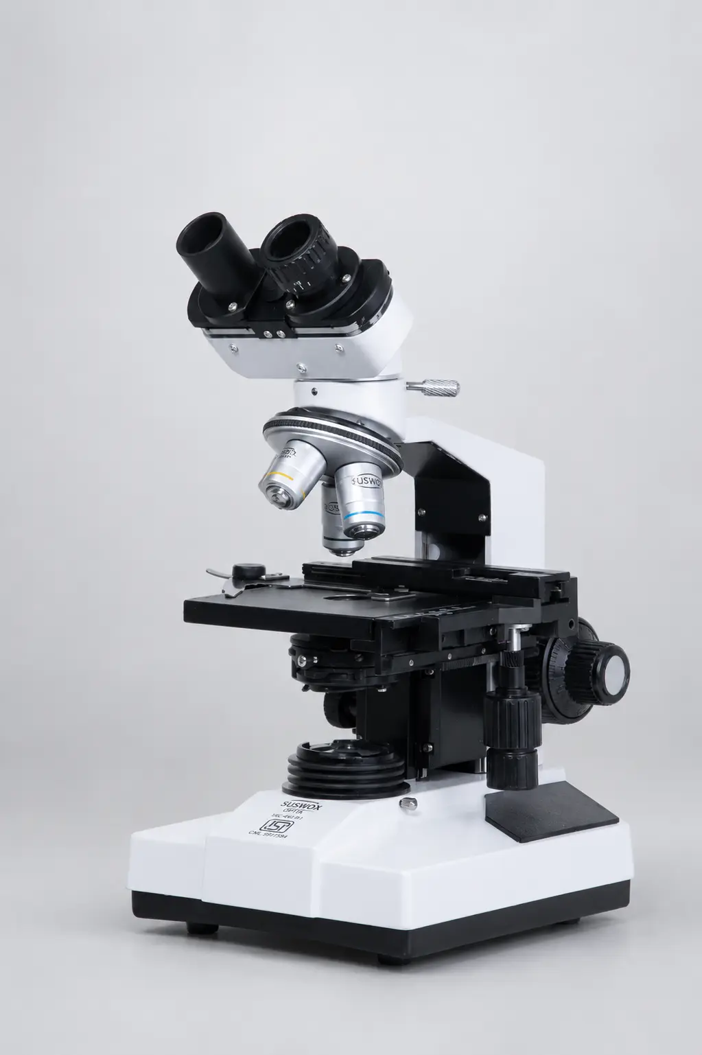
A binocular microscope is a microscope with two eyepieces, so you view the specimen with both eyes at once instead of one. Inside the head, prisms split the light into two paths — one for each eye — which gives a comfortable, natural, slightly three-dimensional image. It magnifies tiny objects like cells, bacteria, and tissues that the naked eye simply cannot see.
And that is why you’ll find binocular microscopes everywhere — schools, pathology labs, hospitals, research centres, and factory quality-control rooms. Both eyes working together means less strain, better depth, and far less fatigue during long sessions.
Simple definition, big difference in real use.
Binocular microscope diagram
Before the parts make sense, it helps to see them. Here is a labelled diagram of a binocular microscope.
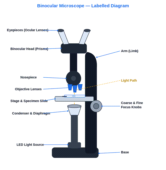
Labelled diagram of a binocular microscope and the path light takes through it.
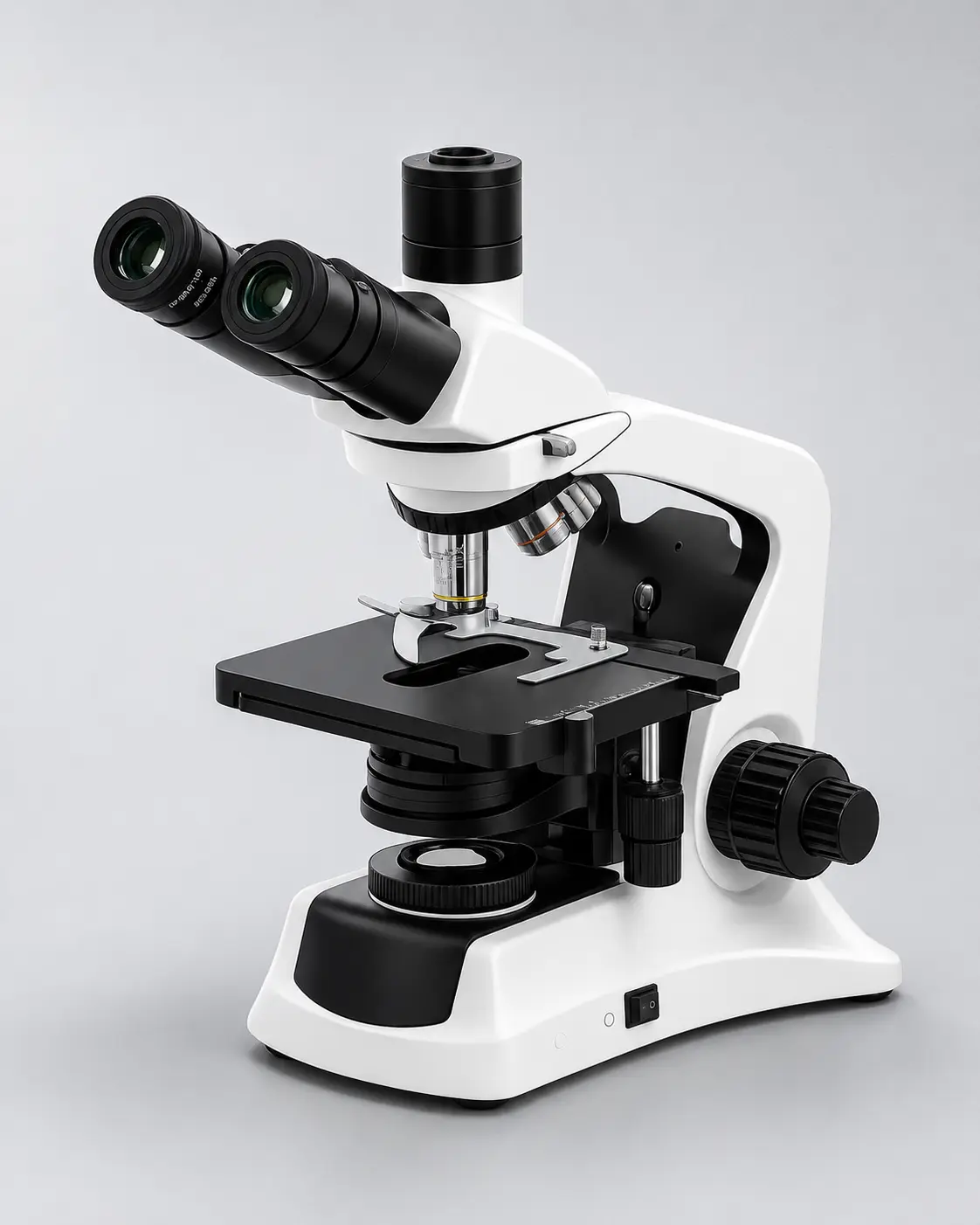
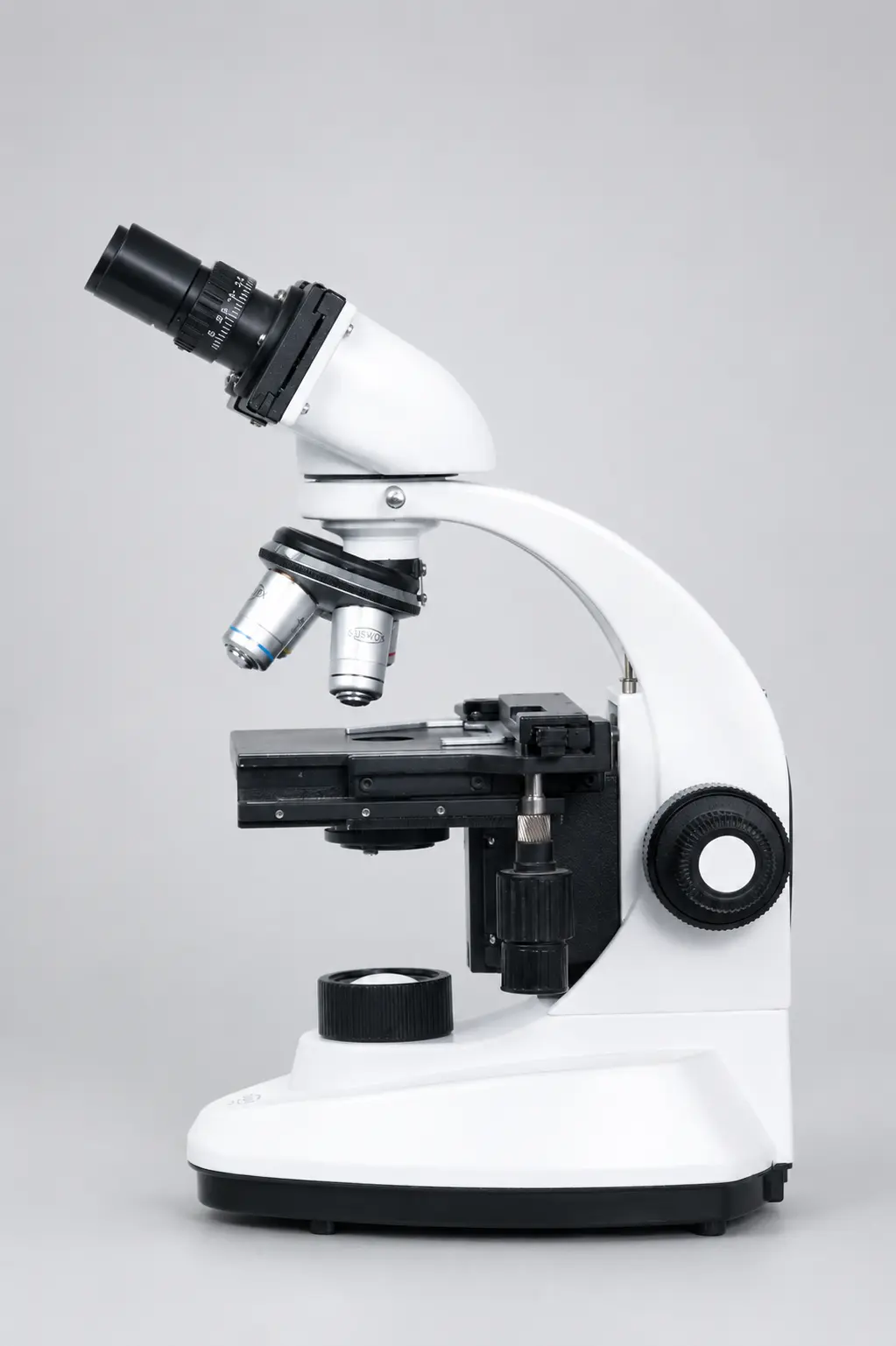
Parts of a binocular microscope
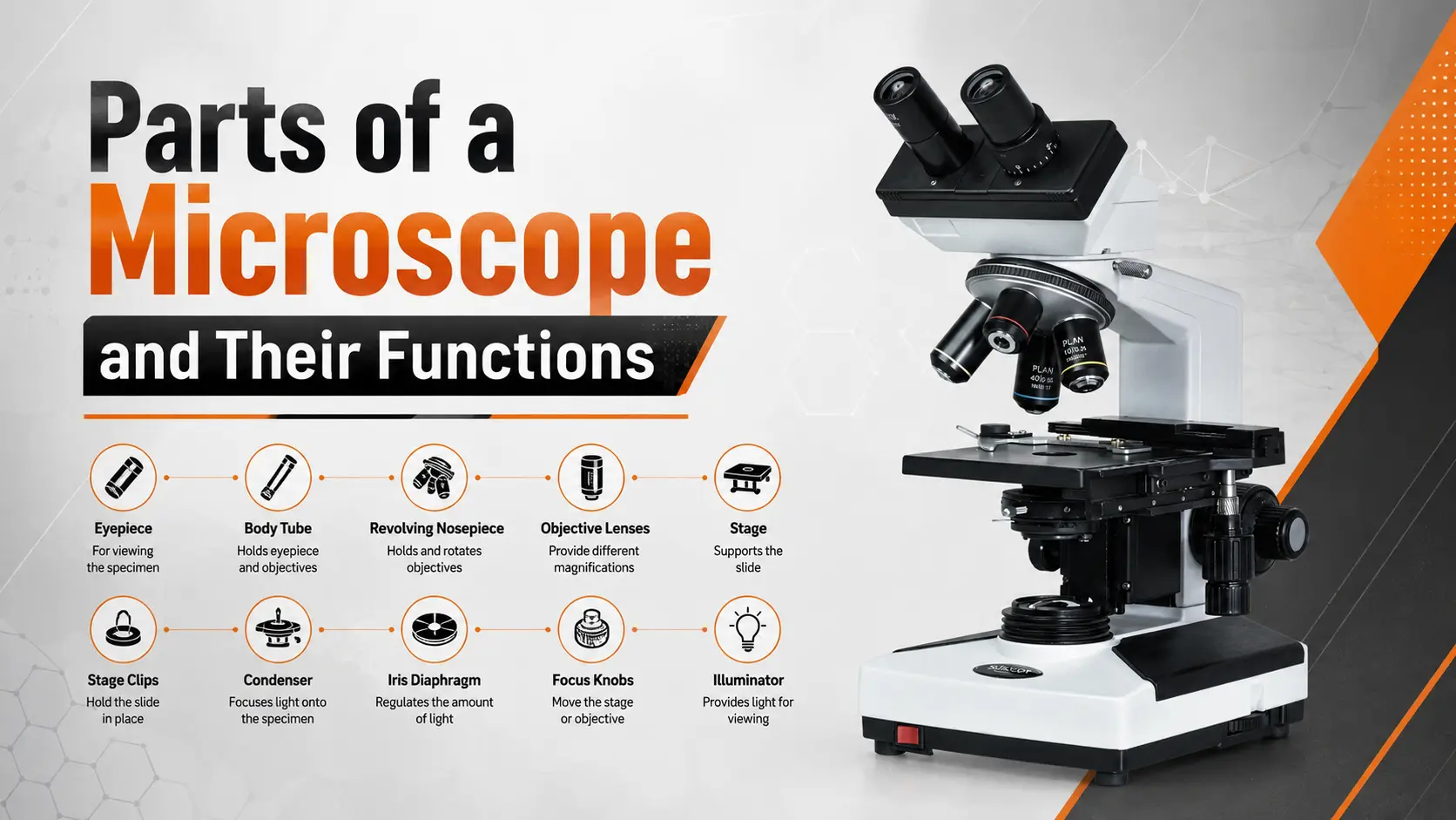
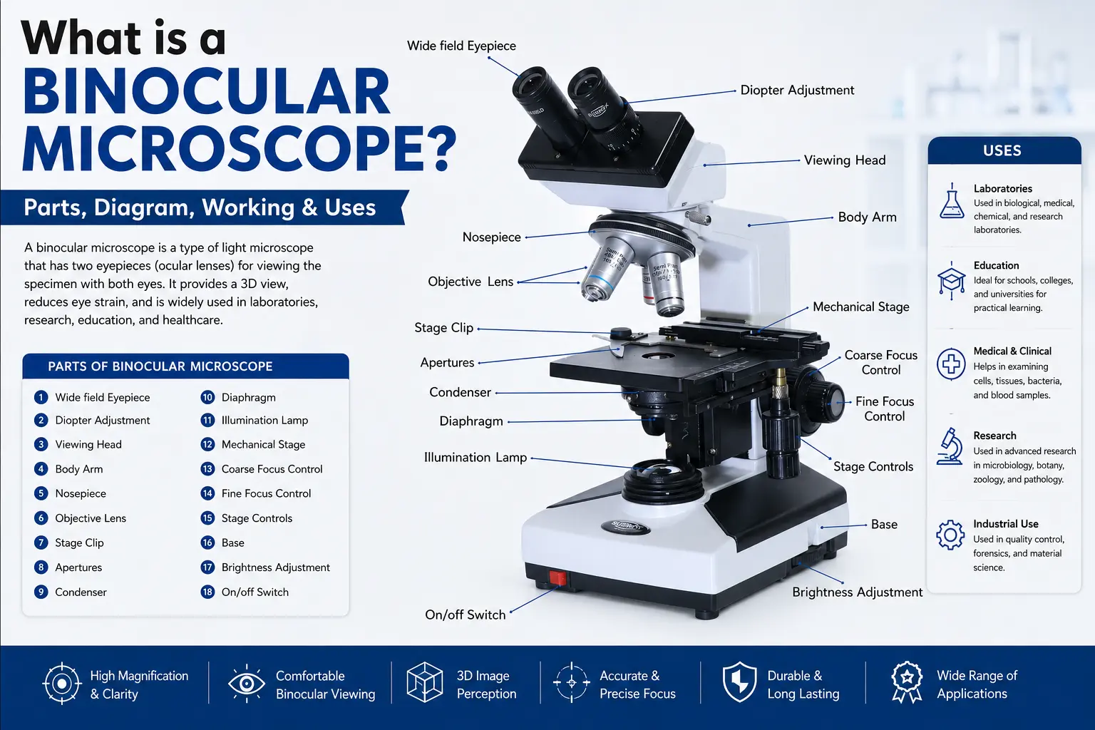
A binocular microscope looks complex, but it is really just a handful of parts working together. Here is what each one does.
| Part | What it does |
| Eyepieces (ocular lenses) | The two lenses you look through — usually 10x or 15x. They magnify the image a second time. |
| Objective lenses | Sit on the nosepiece — normally 4x, 10x, 40x and 100x. They do the main magnifying. |
| Binocular head (prisms) | Splits one image into two paths, one for each eye, to create the 3D effect. |
| Nosepiece | The rotating turret that holds the objective lenses so you can switch magnification. |
| Stage | The flat platform where you place the specimen slide, held by clips. |
| Condenser & diaphragm | Focus the light onto the specimen and control how bright it is. |
| Focus knobs | Coarse knob for rough focus, fine knob for sharp detail. |
| Light source | An LED or halogen lamp that shines light up through the specimen. |
| Arm & base | Hold the whole microscope steady and make it easy to carry. |
The clever bit is the head. In a monocular microscope, one image goes to one eye. Here, prisms take that single image and hand a copy to each eye — which is exactly what makes it so comfortable to use.
How does a binocular microscope work?
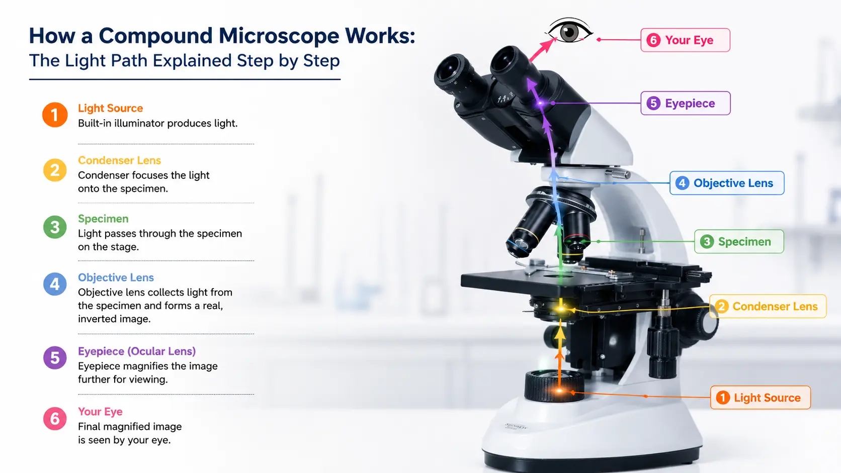
A binocular microscope works by shining light up through the specimen, magnifying it with the objective lens, then using prisms to split that image into two paths so both eyes see it at once. The result is a bright, sharp, slightly 3D view.
Step by step, here is the journey the light takes:
- Light on. The LED or halogen lamp at the base sends light upward.
- Through the condenser. The condenser and diaphragm focus that light and set its brightness.
- Through the specimen. Light passes up through the slide on the stage.
- Into the objective. The objective lens catches the light and magnifies the image.
- Split by prisms. Prisms in the head divide the image into two identical paths.
- To both eyes. Each eyepiece delivers the image to one eye, creating a comfortable, depth-rich view.
No guesswork. Just light, lenses, and prisms doing their job in order.
Types of binocular microscopes
Not every binocular microscope is the same. There are three common types, and picking the right one depends on what you’re looking at.
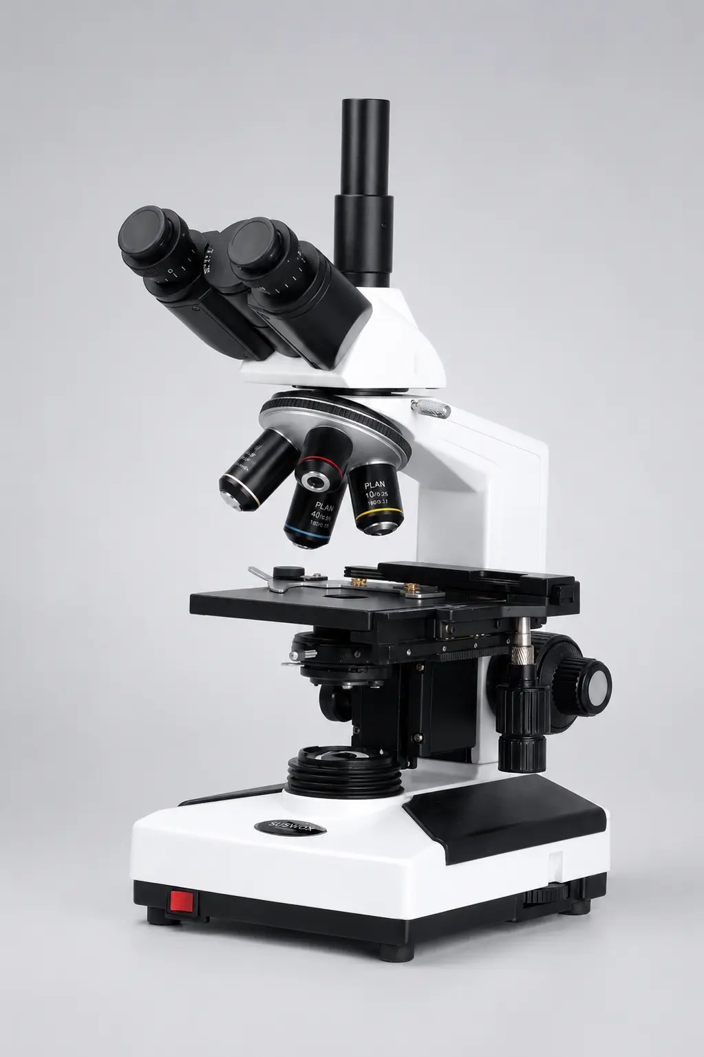
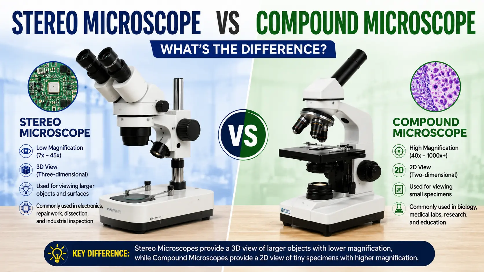
- Compound binocular microscope — for thin, transparent samples like cells and bacteria. High magnification, usually up to 1000x. This is the classic lab and medical microscope.
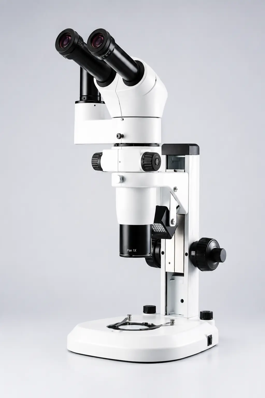
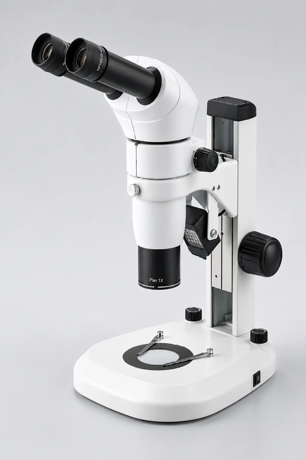
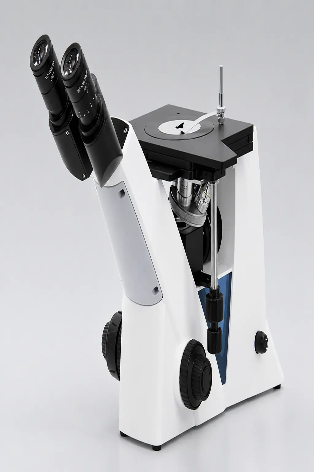
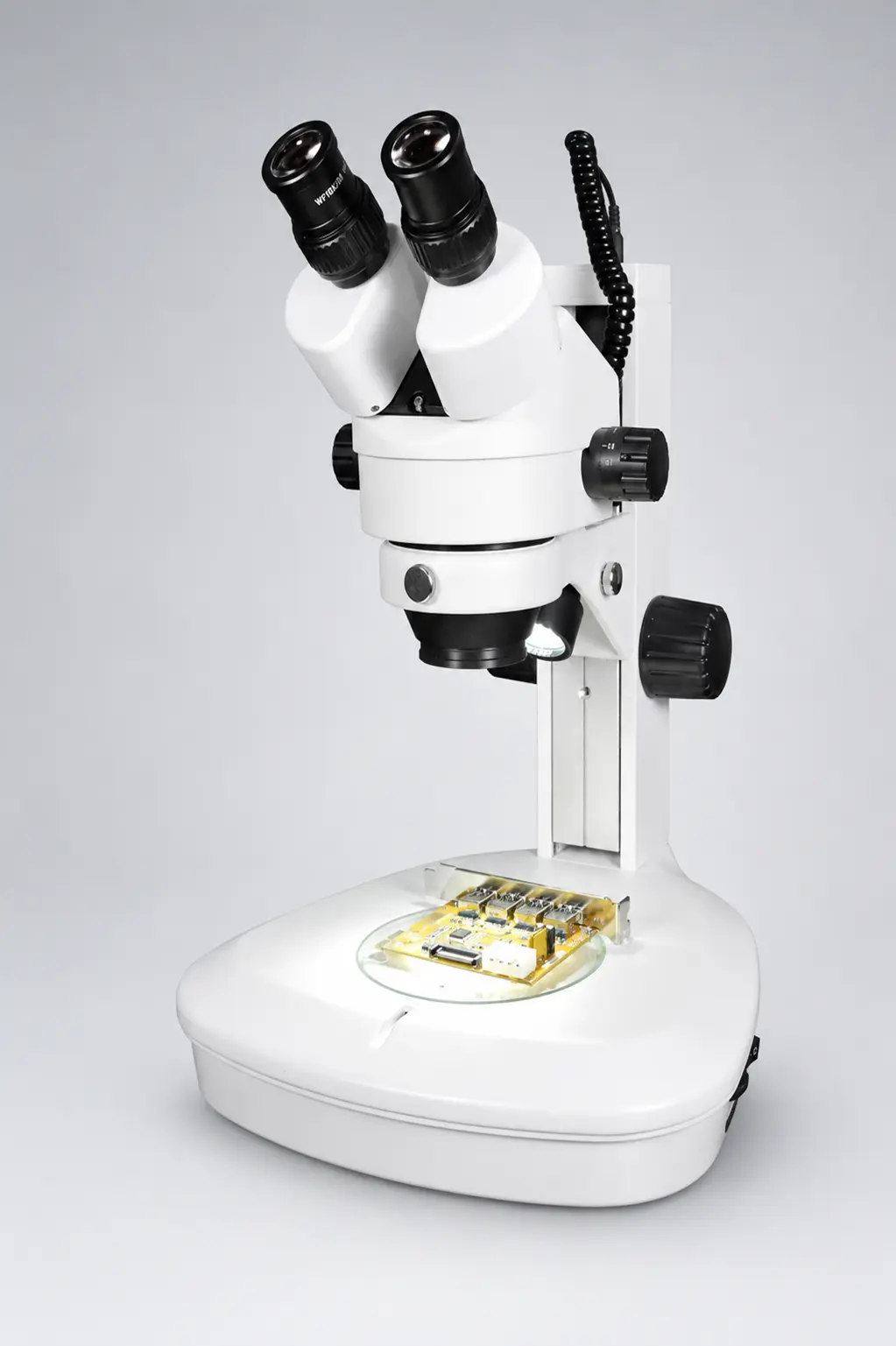
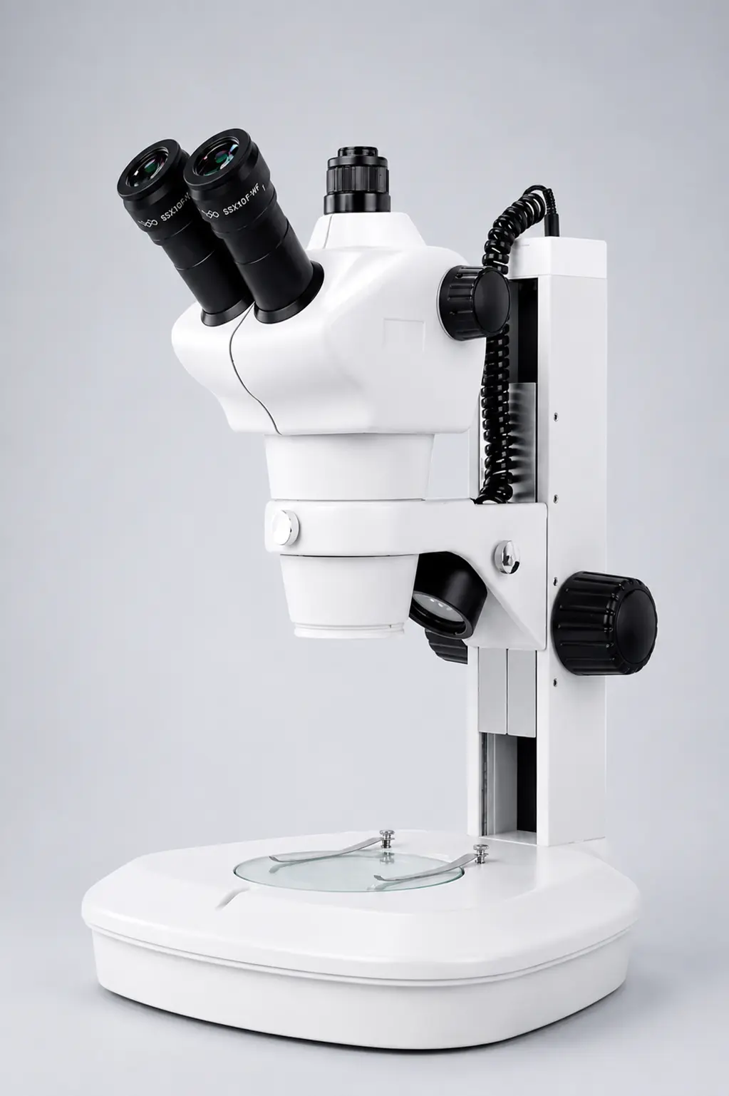
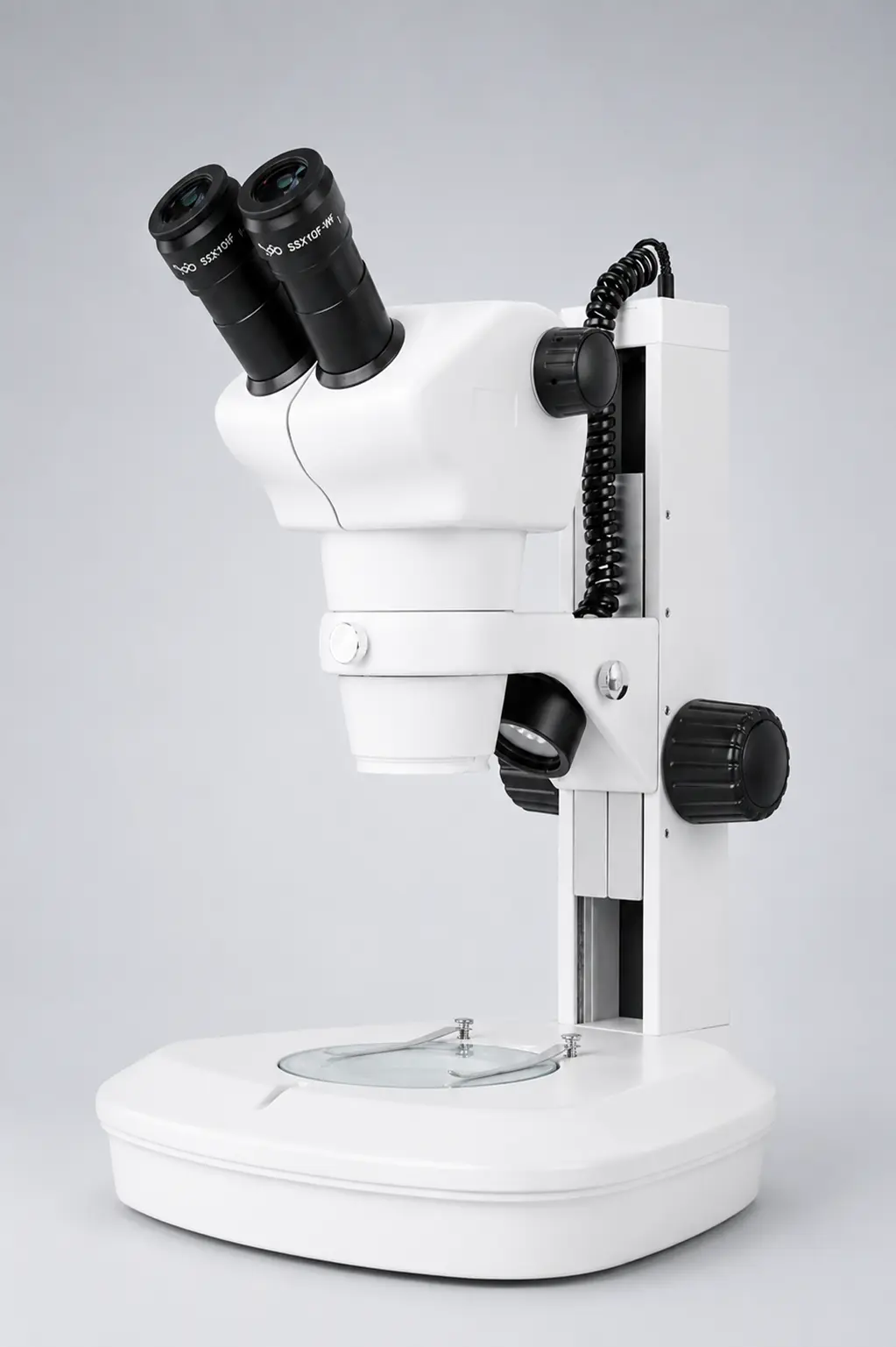
- Stereo (stereoscopic) binocular microscope — for larger, solid objects like insects, plants, coins, and circuit boards. Lower magnification (around 10x–80x) but a strong 3D view, great for dissection and inspection.
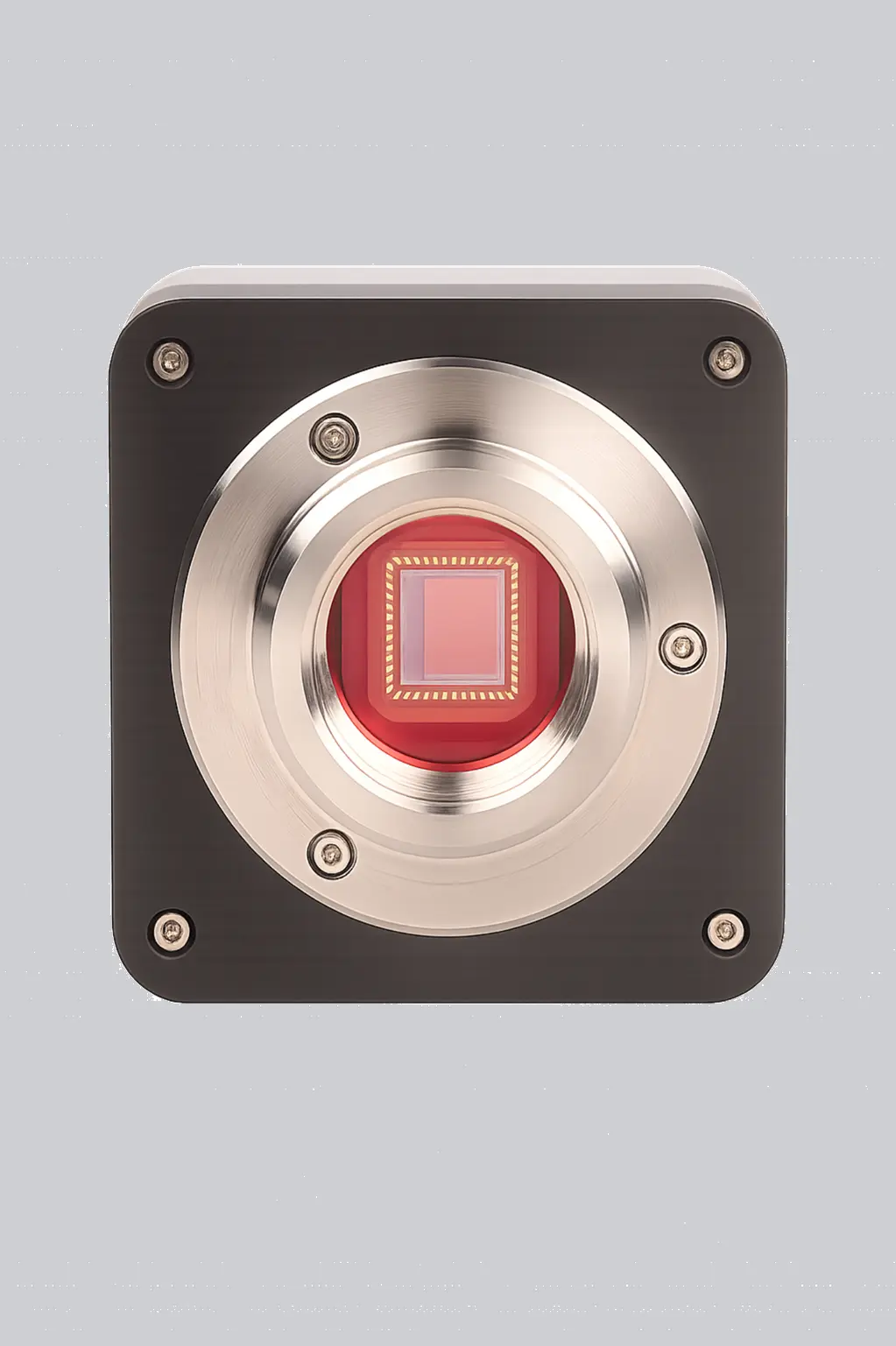
- Digital binocular microscope — an optical microscope with a built-in camera. It shows the image on a screen, which is perfect for teaching, sharing, and recording results.
Binocular vs monocular vs trinocular microscope
People mix these up all the time, so here is a quick, clear comparison.
| Feature | Monocular | Binocular | Trinocular |
| Eyepieces | One | Two | Two + 1 camera port |
| Eye comfort | Lower | High | High |
| Depth / 3D view | Limited | Good | Good |
| Best for | Basic school use | Labs, clinics, long study | Research & imaging |
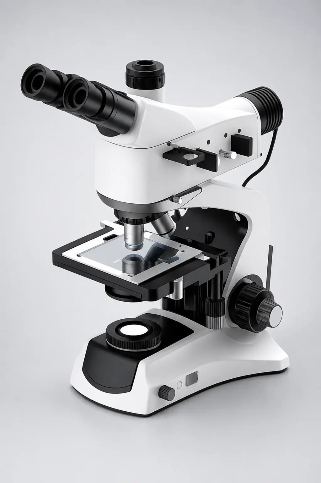
| Photo / video | No | Rarely | Yes (camera ready) |
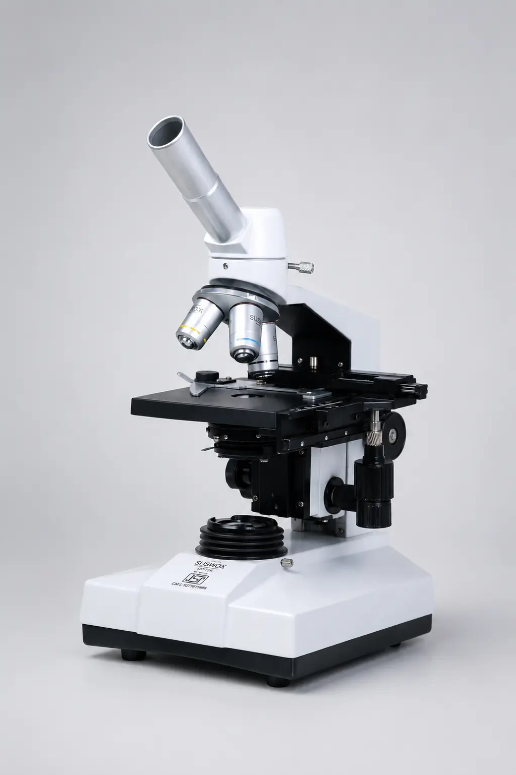
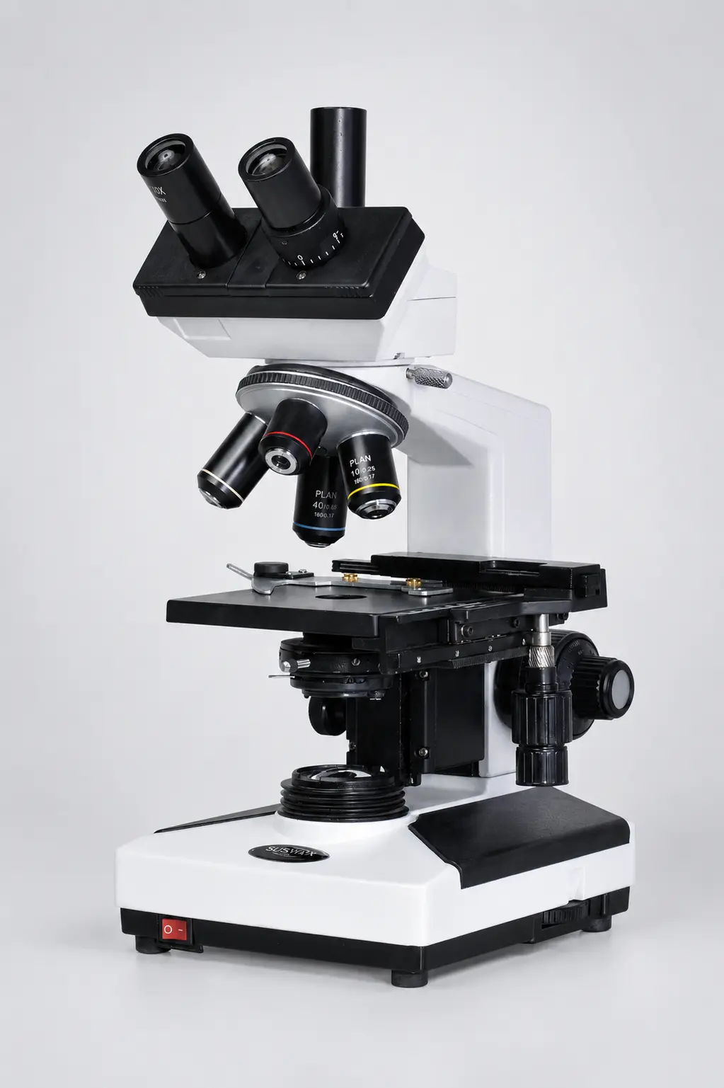
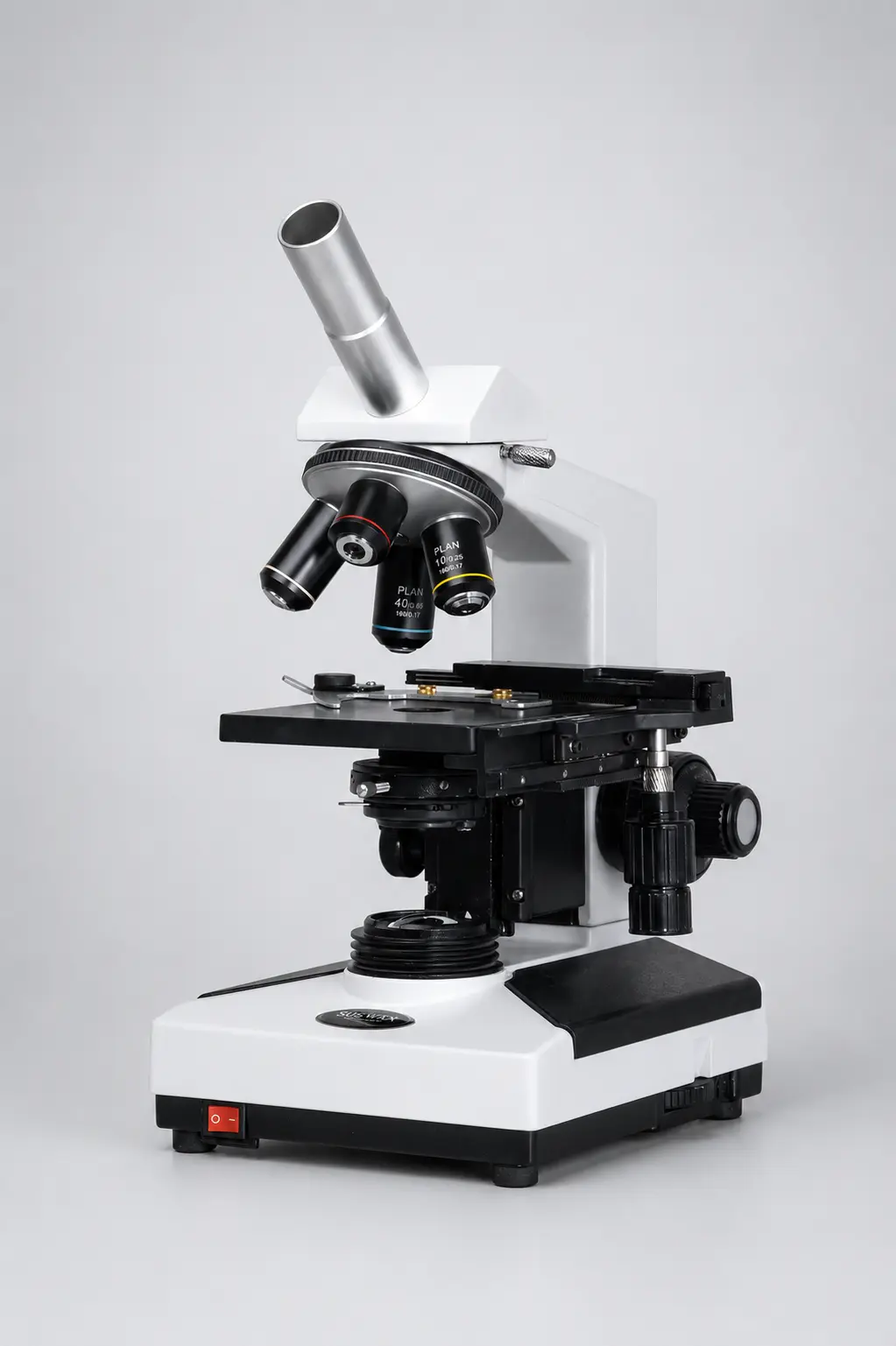
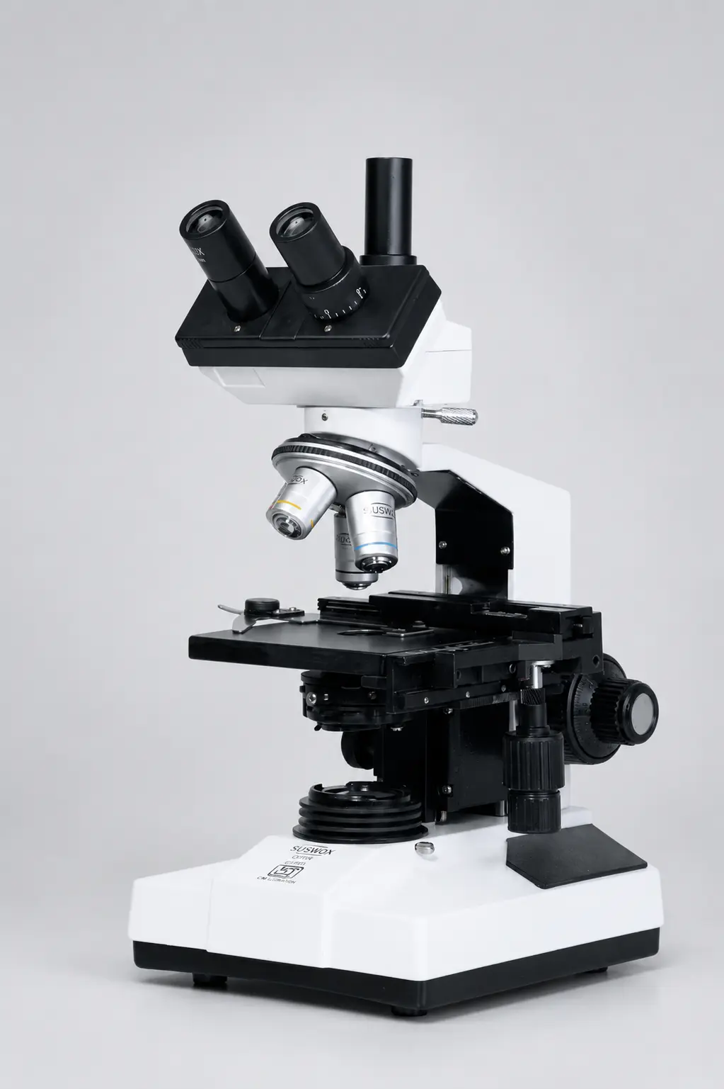
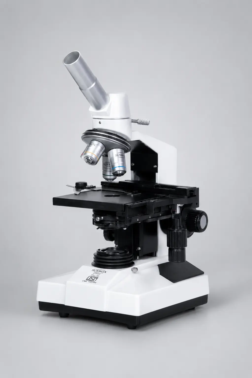
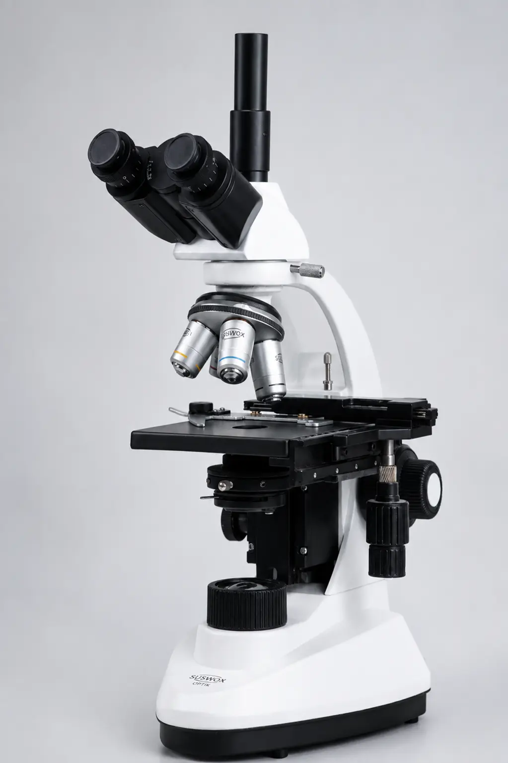
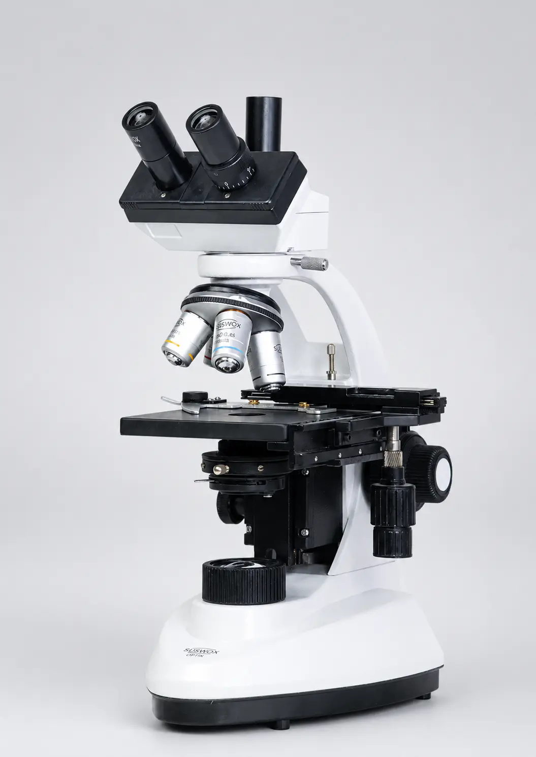
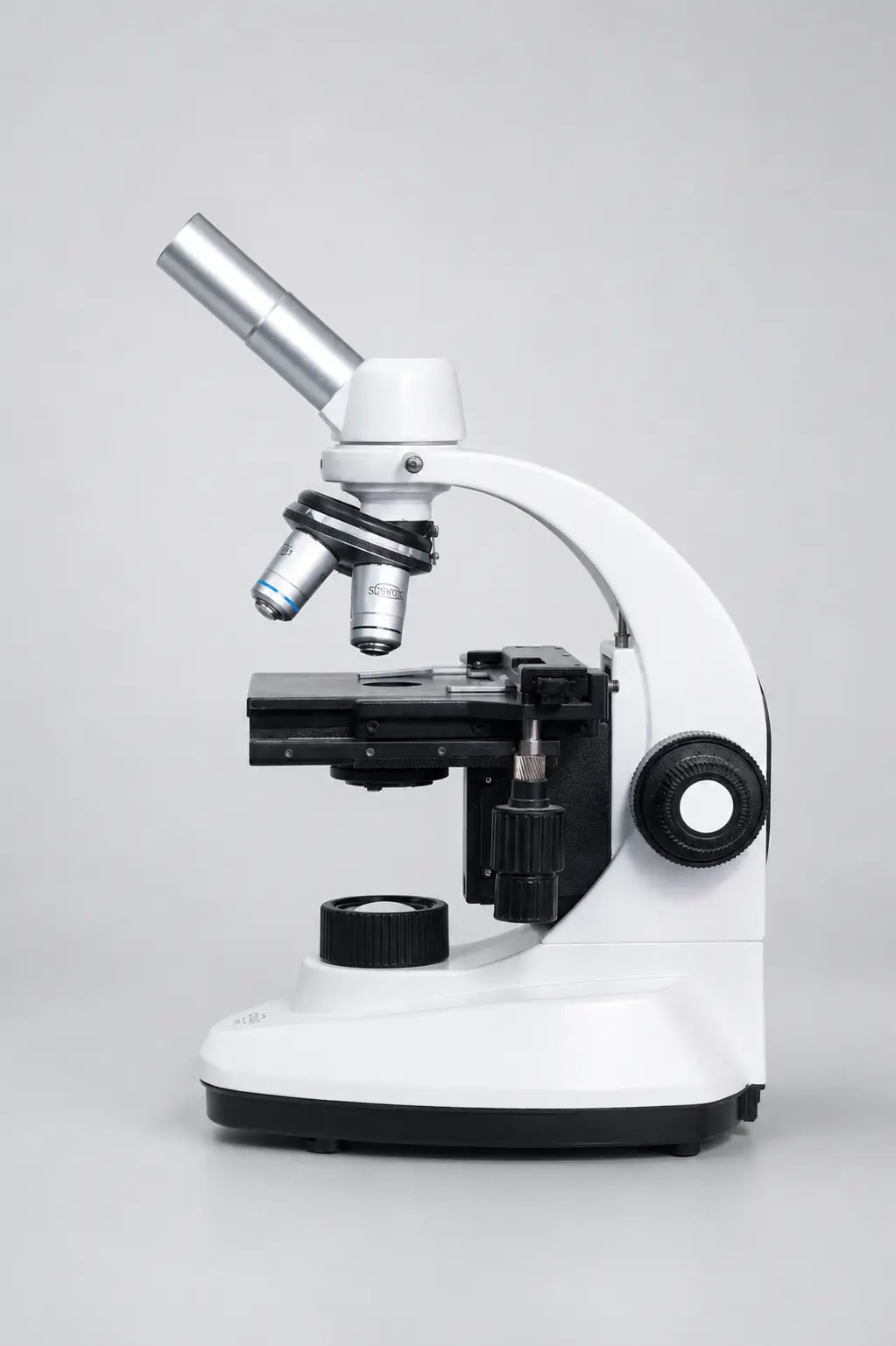
Short version: a monocular has one eyepiece and suits basic school use. A binocular has two and is the comfortable all-rounder for labs and clinics. A trinocular adds a third port for a camera, which research and imaging work needs.
Uses of a binocular microscope
Where does this instrument actually earn its place? Pretty much anywhere small detail matters.
- Education — students study biology, cells, and material science with comfort during long classes.
- Medical laboratories — technicians examine blood, urine, tissues, and microorganisms for diagnosis.
- Research centres — scientists study cell structures, samples, and reactions in detail.
- Industrial inspection — engineers check electronics, metal parts, and precision components for tiny faults.
Benefits of a binocular microscope
Why choose binocular over a single-eyepiece model? A few solid reasons.
- Far less eye strain during long study or lab sessions.
- A more natural, relaxed head and neck position.
- Better depth perception and a slight 3D effect.
- Sharper attention to fine detail without tiring quickly.
- Reliable for both teaching and serious research.
How to choose the right binocular microscope
Buying one? Don’t just look at the price sticker. Check these first.
- Purpose. Compound for biology and cells, stereo for inspection and dissection. Match the tool to the task.
- Magnification. Pick objective and eyepiece combinations that give the detail you actually need.
- Lighting. LED runs cooler and cheaper than halogen, and lasts longer.
- Comfort adjustments. Interpupillary distance and diopter adjustment make long sessions much easier.
- Build quality. A stable base and smooth focus knobs are signs of a microscope that will last.
Simple maintenance tips
Look after it and a good microscope lasts for years.
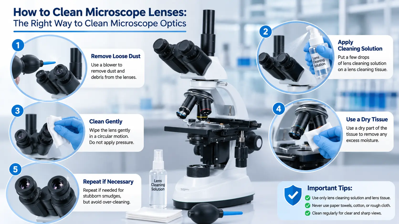
- Never clean lenses with your fingers — use lens paper or a microfiber cloth.
- Cover it with a dust cover when it’s not in use.
- Store it in a dry, dust-free place.
- Switch off the light source when you’re done.
- Check the moving parts now and then so focusing stays smooth.
Frequently Asked Questions
What is a binocular microscope in simple words?
A binocular microscope is a microscope with two eyepieces, so you look through it with both eyes instead of one. This gives a comfortable, natural, slightly 3D view and greatly reduces eye strain compared to a single-eyepiece (monocular) microscope.
What are the main parts of a binocular microscope?
The main parts are two eyepieces (ocular lenses), objective lenses on a rotating nosepiece, the binocular head with prisms, the stage for the slide, a condenser and diaphragm, coarse and fine focus knobs, a light source, and the arm and base for support.
How does a binocular microscope work?
Light shines up from the base through the specimen, the objective lens magnifies the image, and prisms in the head split it into two paths — one for each eye. Both eyes then see the same magnified image, creating a clear, comfortable, three-dimensional view.
What is the difference between a binocular and a monocular microscope?
A binocular microscope has two eyepieces and lets you use both eyes, giving more comfort, better depth, and less fatigue. A monocular microscope has just one eyepiece and is cheaper and simpler, which suits basic school use.
What magnification does a binocular microscope give?
Magnification comes from combining the objective lens (usually 4x, 10x, 40x, or 100x) with the eyepiece (usually 10x). So a compound binocular microscope can reach from about 40x up to 1000x, while a stereo model gives lower magnification, around 10x to 80x.
Where are binocular microscopes used?
They are used in schools and colleges for teaching, in medical and pathology labs for diagnosis, in research centres for studying cells and samples, and in industry for inspecting electronics and precision parts.
Final thoughts
So — what is a binocular microscope? It’s the comfortable, two-eyed workhorse of the microscope world. Same magnifying power as a single-eyepiece model, but far easier on your eyes and much better at showing depth. Whether you’re a student, a lab technician, or a researcher, it’s the tool that lets you look closely for hours without paying for it later with a headache.
Understand how it works, keep it clean, and it will serve you for years.
Looking to buy a binocular microscope?
Suswox builds ISI-marked binocular, pathological, research, and stereo microscopes trusted by labs, schools, and universities since 1973. As a leading Microscope Manufacturer & Exporter in India, Suswox delivers research-grade clarity at honest, Indian-made prices — with local service and spares always close by. Explore the full range at suswox.com.